Clear Sky Science · en
Regionalized regulation of actomyosin organization influences cardiomyocyte cell shape changes during chamber curvature formation
How Heart Cells Sculpt the Organ’s Curves
Our hearts are not simple pumps; they are precisely shaped machines whose curves help direct blood efficiently. This study asks a deceptively simple question: how do individual heart muscle cells change their shapes to carve the bulges and bends of a working heart? By zooming in on tiny cells in the embryonic zebrafish heart, the authors reveal how an internal “muscle scaffold” in each cell is tuned differently in neighboring regions, helping to bend a straight heart tube into a fully formed chamber.
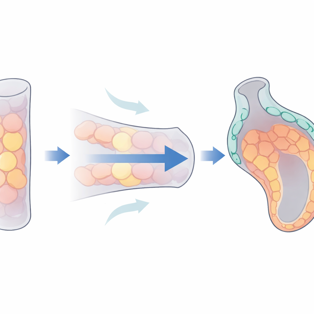
From Straight Tube to Curved Heart
In vertebrate embryos, the heart begins as a narrow tube that later loops and balloons out to form separate chambers. Each chamber develops two distinct regions: a bulging outer curvature and a recessed inner curvature. These regions do not just look different at the tissue level; they also beat differently and have distinct stiffness and internal structure. Yet, the steps that first distinguish outer and inner regions, and how those differences emerge from the behavior of single cells, have been unclear. Zebrafish, whose transparent embryos allow live imaging of the beating heart, offer an ideal system to track these events in space and time.
Heart Cells Stretch Out or Stand Tall
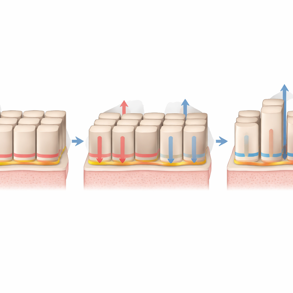
The researchers first followed how heart muscle cells (cardiomyocytes) in the nascent ventricle change shape as the chamber curves. Early on, cells in what will become the outer and inner curvatures are nearly identical in size and outline. As development proceeds, both sets of cells grow, but they deploy that extra volume differently. Outer curvature cells spread mainly in the plane of the heart wall, becoming thin and squamous, like paving stones laid side-by-side. Inner curvature cells, by contrast, elongate mainly from the inner to outer surface, becoming more cuboidal or column-like. These differences arise while the heart is still relatively tubular, pointing to active, region-specific cell shape changes as a driver—rather than just a consequence—of chamber curvature.
The Cell’s Inner Scaffold Sets the Tone
To uncover what orchestrates these contrasting shapes, the team focused on actomyosin, a network of protein filaments that can both pull and push on cell membranes. At early stages, cells destined for outer and inner curvatures show similar distributions of this scaffold. But around the time curvature begins, a striking pattern emerges: in outer curvature cells, actomyosin becomes enriched along the basal side—the surface contacting the surrounding matrix—while inner curvature cells accumulate more scaffold along their lateral and apical surfaces. This shift in internal architecture precedes the visible shape differences, and when the authors used drugs or genetic tricks to dampen actomyosin activity, outer curvature cells failed to spread into their normal thin form and instead resembled the taller inner cells. Mosaic experiments, where only some cells in the heart had reduced actomyosin function, showed that each cell’s own scaffold is crucial: cells with impaired actomyosin stayed stubby even when surrounded by normal neighbors.
Forces from Blood and Genetic Programs Work Together
The heart does not remodel in isolation; it is already pumping blood while it forms. The study shows that blood flow itself helps tune the actomyosin scaffold. In zebrafish mutants with weak atrial contraction and reduced flow through the ventricle, outer curvature cells neither enriched their basal scaffold nor flattened properly. Their internal filaments shifted toward the sides and top of the cell, and the cells elongated in the wrong direction. Intrinsic genetic programs also matter. When the authors disrupted tbx5a—a gene known to control many outer curvature–specific features—outer curvature cells again lost their basal scaffold bias and failed to spread in the plane of the wall. Transplant experiments, where wild-type and tbx5a-deficient cells were mixed in the same heart, revealed that tbx5a acts partly within each cell, but the surrounding tissue environment can modulate its impact.
How Microscopic Changes Shape a Beating Organ
Taken together, the work outlines a clear chain of events for chamber shaping. Blood flow and gene activity converge to rearrange the actomyosin scaffold inside outer curvature heart cells, concentrating it at the base where cells contact their matrix. This configuration appears to let cells push their basal surface outward and spread sideways, while keeping tension low at the top surface so it can expand passively. Inner curvature cells, with more scaffold toward their apical and lateral sides and less at the base, tend to grow upward rather than outward. Through these coordinated, region-specific choices in cell architecture and shape, a straight embryonic heart tube is sculpted into a chamber with a bulging outer wall and recessed inner wall—geometry that is essential for robust heart function.
Citation: Leerberg, D.M., Avillion, G.B., Priya, R. et al. Regionalized regulation of actomyosin organization influences cardiomyocyte cell shape changes during chamber curvature formation. Nat Commun 17, 3768 (2026). https://doi.org/10.1038/s41467-026-70384-5
Keywords: heart development, cell shape, cytoskeleton, zebrafish, biomechanics