Clear Sky Science · en
A topographical organization in the primary olfactory cortex
The Hidden Map Behind Our Sense of Smell
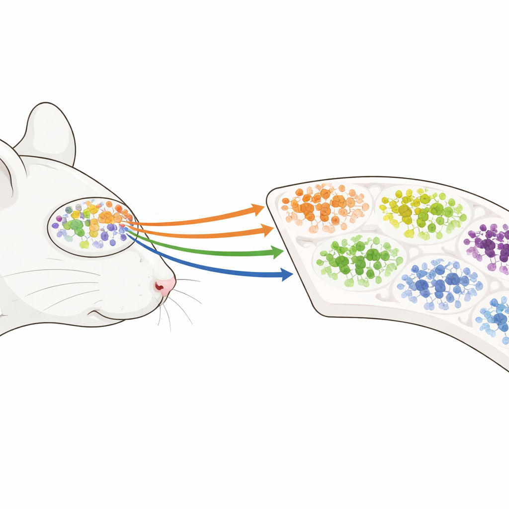
We usually think of smell as something messy and mysterious: countless odor molecules swirling into the nose and somehow becoming distinct scents in our minds. In vision and touch, scientists have long known that the brain uses orderly maps, where neighboring brain cells respond to neighboring spots in space. This study asks whether something similar, a hidden map, also exists for smell in a part of the brain called the primary olfactory cortex, and reveals that such an organized layout does in fact exist.
From Nose Surface to Brain Circuits
Odor molecules entering the nose first activate structures called glomeruli in the olfactory bulb, the brain’s first relay for smell. Each glomerulus responds to certain types of odor molecules. For vision or touch, nearby points on the eye or skin are wired to nearby brain cells, creating orderly maps. But decades of work suggested that smell might be different, with connections from bulb to cortex seemingly jumbled and random. The authors revisited this puzzle with new tools, asking not just where inputs came from, but how groups of glomeruli together drive individual neurons deeper in the brain.
Lighting Up Smell Pathways With Tiny Patterns
To uncover this organization, the researchers turned the olfactory bulb into a controllable “input screen.” Using mice whose bulb neurons could be activated by light, they projected thousands of small blue light patterns across the bulb’s surface while recording the electrical activity of many neurons in the anterior piriform cortex, a key smell-processing area. By tracking which bulb locations had to be lit, alone or in combination, to make a given cortical neuron fire, they built a kind of receptive field map for each cortical cell: a list of glomeruli that excited it, inhibited it, and how strongly.
Many Inputs, Flexible Responses
The maps showed that a typical cortical neuron draws on signals from several dozen glomeruli spread across the bulb, once the full bulb is taken into account. Some of these inputs push the neuron to fire, others hold it back. Importantly, a neuron did not act like a detector for one precise pattern of bulb activity. Instead, it could be activated by several different small subsets of its input glomeruli, as long as the overall incoming drive was strong enough. In other words, the same neuron could respond to multiple distinct combinations of smell signals, suggesting a flexible, overlapping code rather than a rigid “one pattern, one neuron” scheme.
Nearby Neurons Share More of the Same Smell Inputs
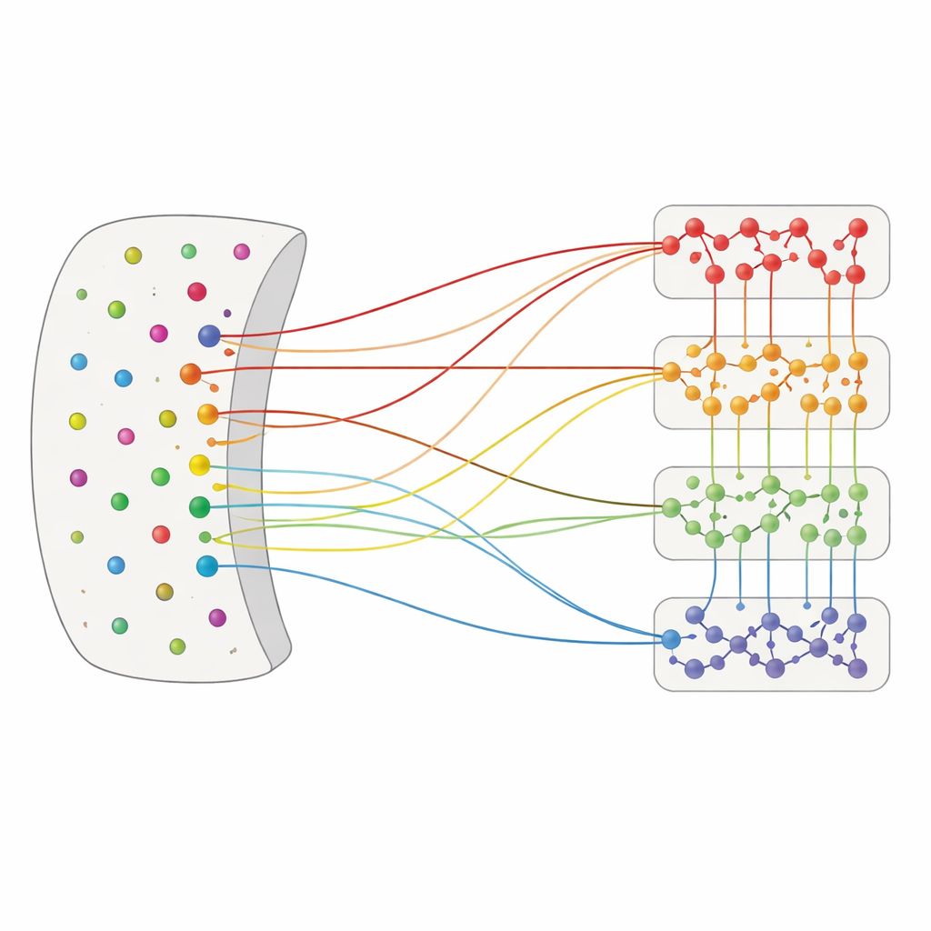
When the team compared the input maps of many cortical neurons, a clear trend emerged: neurons sitting close together in the primary olfactory cortex tended to draw on more similar sets of glomeruli than neurons that were farther apart. Their preferred glomeruli often lay near one another on the bulb, and neighboring neurons sometimes even shared specific glomeruli, though with excitatory or inhibitory effects that could differ. As the physical distance between two cortical neurons grew, the similarity between their input maps declined. This pattern held up across many recording sessions and careful checks that separate neurons were not being confused with each other.
Input Maps Match How Neurons Respond to Real Odors
The researchers then asked whether this wiring pattern shows up in how neurons respond to actual smells. By analyzing both new data and previously published recordings of cortical responses to panels of odors, they found that neurons located near one another tended to have slightly more similar odor preferences than distant ones. The effect was small but consistent in both awake and anesthetized animals. Moreover, pairs of neurons with more similar input maps from the bulb also tended to respond more similarly to odor blends, linking the hidden wiring diagram directly to real sensory behavior in the cortex.
What This Means for How We Smell
For years, olfactory cortex was thought to be largely unstructured, a tangle of random connections unlike the neat maps seen in other senses. This work shows that, beneath the apparent randomness, there is a subtle topographical rule: neurons that sit close together in the primary olfactory cortex tend to receive more similar mixtures of smell information from the nose and respond to odors in more similar ways. Rather than a simple one-to-one map, smell uses a many-to-one pattern in which overlapping groups of glomeruli are funneled into clusters of nearby cortical neurons. This organization may help the brain balance wiring cost, flexibility, and the ability to recognize related smells while still distinguishing among them.
Citation: Taragin, S., Bashan, O., Dalal, T. et al. A topographical organization in the primary olfactory cortex. Nat Commun 17, 3994 (2026). https://doi.org/10.1038/s41467-026-70356-9
Keywords: olfactory cortex, sensory maps, neural circuits, odor coding, topographic organization