Clear Sky Science · en
Purkinje cell intrinsic activity shapes cerebellar development and function
How Early Brain Rhythms Shape Balance and Coordination
Learning to sit, stand, and walk may feel effortless in early life, but beneath those milestones lies a finely tuned “timing center” in the back of the brain: the cerebellum. This study reveals that tiny nerve cells inside the cerebellum—called Purkinje cells—must generate their own steady electrical rhythms very early in life for the brain’s movement circuits to wire up correctly. When those inner rhythms are dampened in newborn mice, their brain circuits develop abnormally and the animals grow up with poor balance and clumsy, uncoordinated movements. 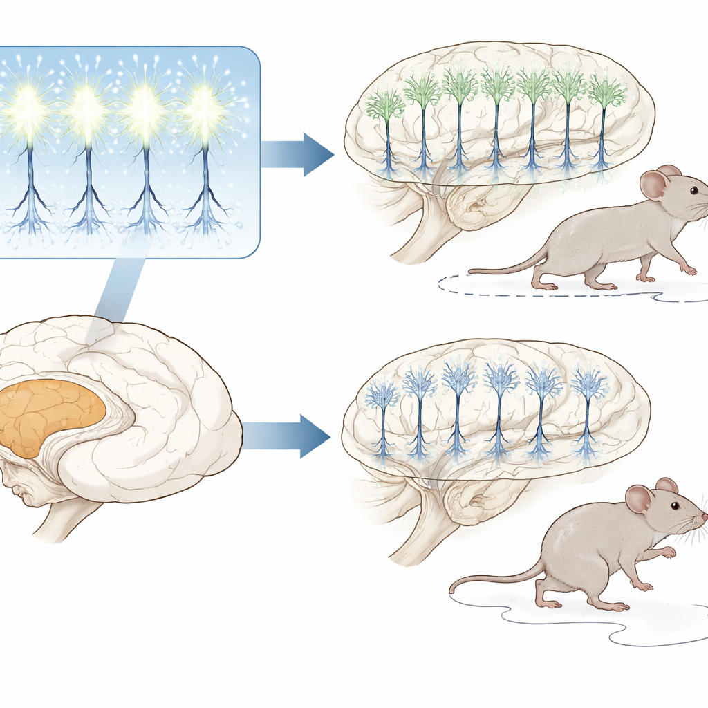
Key Players in the Brain’s Movement Control Center
The cerebellum helps us keep our balance, coordinate our limbs, and fine‑tune movements like tracking a moving object with our eyes. Purkinje cells sit at the heart of this system. They are large, branching neurons that constantly send signals to deeper cerebellar nuclei, which in turn talk to the rest of the brain and spinal cord. Remarkably, Purkinje cells can fire electrical impulses on their own, even before they receive many inputs from other neurons. This built‑in activity, or intrinsic firing, has long been suspected to guide how cerebellar circuits form, but its exact role during early development has been unclear.
Turning Down Purkinje Cell Activity in Young Brains
To test how crucial these early rhythms are, the researchers engineered mice in which Purkinje cells could be selectively “quieted” at chosen ages. They used a genetic switch to boost a potassium channel that holds the neuron’s electrical voltage more negative, making spontaneous firing far less likely. This manipulation strongly reduced Purkinje cell firing both in brain slices and in awake animals, yet the cells could still respond when artificially stimulated. By flipping this switch during different postnatal weeks, the team could compare what happens when intrinsic activity is disrupted from birth, from the second week, or later in development.
How Silenced Cells Distort Brain Wiring
When Purkinje cell activity was suppressed from the first days of life, their growth and shape were dramatically altered. Normally, each Purkinje cell develops a single, broad, fan‑like tree of branches that reaches into the upper cerebellar layer. In activity‑reduced mice, these trees were smaller, less complex, and extended less far. The cells also formed fewer inhibitory contacts onto neurons in the deep cerebellar nuclei, which then fired more erratically. Microscopic analyses showed that incoming connections from other pathways—climbing fibers and parallel fibers—were also miswired or stunted. Together, these changes indicate that early Purkinje firing instructs both the growth of their own branches and the precision of the connections they make and receive. 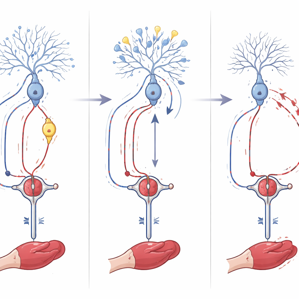
From Miswired Circuits to Clumsy Movement
These wiring defects translated into clear movement problems. Adult mice whose Purkinje cells had been silenced from birth walked with an unsteady, ataxia‑like gait, slipped more often on a narrow beam, and fell sooner from a rotating rod. They also struggled with cerebellum‑dependent learning tasks: adapting eye movements to moving surroundings and learning to blink in anticipation of an air puff. Strikingly, when the researchers delayed the onset of Purkinje cell silencing by one or two weeks, the motor problems were milder, and structural defects were less severe. This points to a sensitive early window—roughly the first two postnatal weeks in mice—when intrinsic Purkinje activity is especially vital for setting up accurate motor circuits.
Gene Programs Linking Early Activity to Disease
To uncover how intrinsic firing translates into long‑term changes, the team analyzed which genes were turned on or off in Purkinje cells at one week of age when activity was suppressed. Hundreds of genes involved in synaptic communication, calcium handling, and neuron development were altered. Notably, several of these genes have been linked to human movement disorders and degenerative ataxias. Two in particular, Prkcg and Car8, stood out. By selectively reducing these genes in Purkinje cells, the researchers showed that each helps shape early dendritic growth in opposite ways—one restraining overgrowth, the other promoting proper maturation—supporting the idea that early electrical activity steers development through specific gene networks.
Why This Matters for Human Brain Health
The study concludes that the cerebellum’s main output cells must be electrically active during a brief, early window for the brain’s balance and coordination circuits to assemble correctly. When these intrinsic rhythms are dampened, Purkinje cells grow incorrectly, form faulty connections, and trigger long‑lasting problems in movement and motor learning. Because many of the affected genes are also implicated in human cerebellar disorders, the work suggests that some adult movement diseases may trace back to subtle disruptions in early brain activity. Understanding these early requirements could ultimately guide new strategies to protect or repair cerebellar function in at‑risk infants and in people with inherited ataxias.
Citation: Osório, C., White, J.J., Torrents-Solé, P. et al. Purkinje cell intrinsic activity shapes cerebellar development and function. Nat Commun 17, 3688 (2026). https://doi.org/10.1038/s41467-026-70355-w
Keywords: cerebellum, Purkinje cells, motor coordination, neuronal development, ataxia