Clear Sky Science · en
Multimodal evidence for hippocampal engagement and modulation by functional connectivity-guided parietal TMS
Why this brain study matters to everyday life
Our ability to remember events, learn new information, and regulate emotions depends heavily on a small, deeply buried brain structure called the hippocampus. When this area falters, problems like Alzheimer’s disease, post-traumatic stress, and other memory disorders can emerge. Directly stimulating the hippocampus usually requires brain surgery, which is not practical for most people. This study explores whether a noninvasive method—magnetic pulses delivered from outside the skull—can be steered in a smart, personalized way to influence hippocampal activity safely and reliably.
Using brain wiring maps to guide stimulation
The researchers focused on transcranial magnetic stimulation (TMS), which uses brief magnetic pulses applied to the scalp to nudge brain cells. Although TMS mainly reaches the brain’s surface, signals can travel along existing wiring to deeper regions. The team used functional connectivity, a kind of “traffic map” built from brain scans that shows which regions naturally fluctuate together, to pick the best spot on the parietal lobe—a surface area linked to the hippocampus. In some patients, the parietal target was chosen for its strongest connection to the hippocampus; in others, it was picked without this guidance or aimed at different targets. By comparing these strategies, the scientists asked: does carefully choosing a parietal site based on its connection pattern make TMS more effective at reaching the hippocampus?
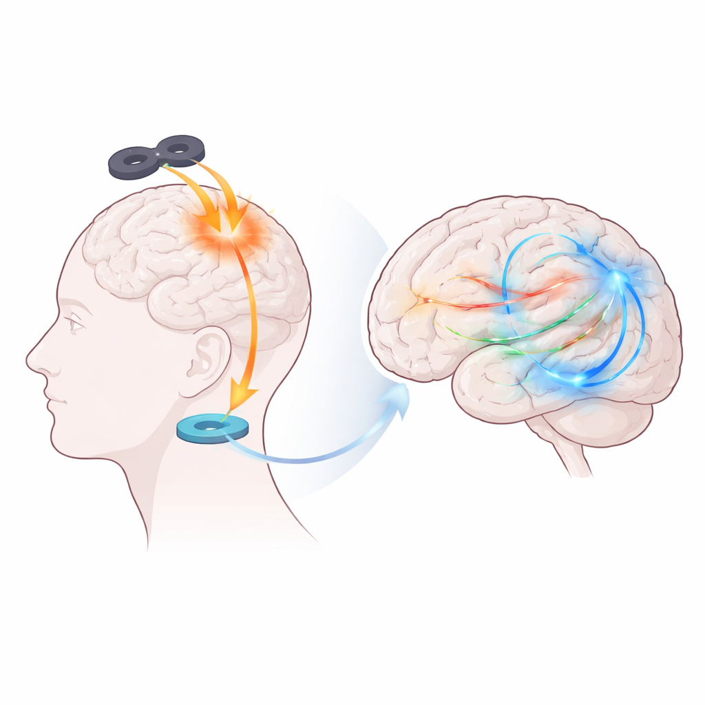
Listening directly to the hippocampus in patients
To obtain direct evidence, the team worked with neurosurgical patients who already had tiny electrodes placed in their brains to monitor epilepsy. In the first experiment, they delivered single TMS pulses over the parietal target while recording electrical activity from the hippocampus. When stimulation sites were selected using the connectivity-guided approach, nearly half of the hippocampal recording contacts showed strong, rapid responses to real TMS but not to sham (placebo-like) pulses. These responses unfolded over several hundredths of a second in distinct time windows and were especially prominent in the theta frequency range—a slow brain rhythm that is a hallmark of hippocampal involvement in memory. In contrast, when the parietal site was not chosen based on hippocampal connectivity, the hippocampus responded far less often, indicating that personalized targeting substantially sharpened engagement of this deep structure.
Mapping person-to-person differences with brain scanning
The second experiment extended these findings to 79 healthy volunteers using brain imaging. Here, participants received single TMS pulses while lying in an MRI scanner, allowing the team to see how blood flow in the hippocampus changed after each pulse. The parietal site used in this dataset had been chosen for other reasons, not specifically for its link to the hippocampus. Even so, individuals varied widely in how strongly that parietal region was functionally connected to their hippocampi at rest. Those with stronger positive connectivity showed larger hippocampal responses to parietal TMS, while those whose wiring placed the regions further apart functionally showed weaker or even negative responses. The closer the actual stimulation site lay to each person’s “optimal” connectivity-defined parietal spot, the stronger the hippocampal response. This supports the idea that a person’s unique connectivity pattern can predict how well surface stimulation will reach deep targets.
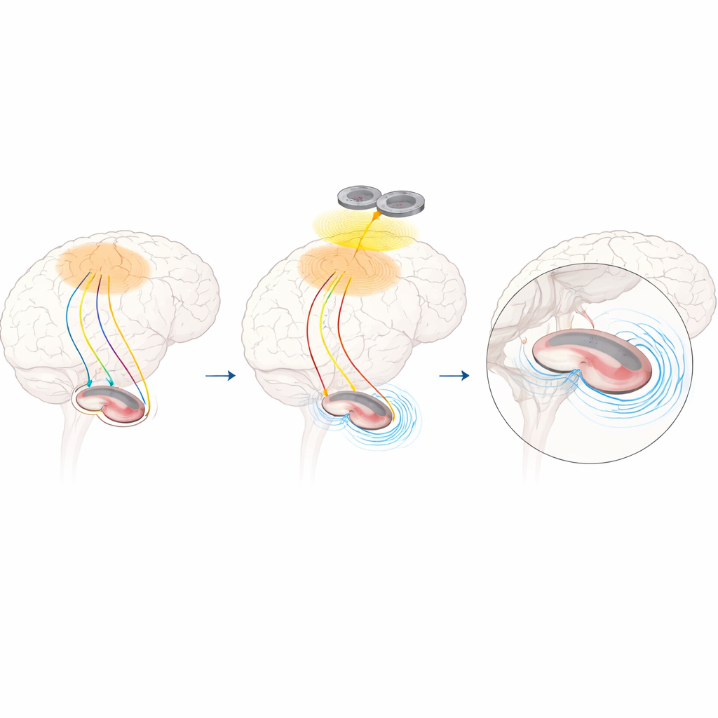
Shaping hippocampal rhythms with repeated pulses
In a third experiment, the researchers asked whether repetitive TMS (rTMS) could not only trigger brief reactions but also reshape ongoing hippocampal rhythms. A subset of neurosurgical patients received short trains of higher-frequency TMS pulses to either connectivity-guided or non-guided parietal sites while hippocampal activity was again recorded directly. When stimulation was guided by hippocampal connectivity, repeated trains produced a robust and lasting reduction in theta power in the hippocampus, building up over successive trains and persisting for more than 20 seconds after each one. This effect was specific: it was much weaker or absent when the parietal site was not chosen based on hippocampal connectivity, and it did not appear to the same degree in neighboring brain regions.
What this means for future treatments
Taken together, these experiments show that noninvasive magnetic stimulation applied to the scalp can causally engage and modulate the hippocampus when it is carefully aimed using individualized connectivity maps. The work offers a mechanistic bridge between earlier behavioral studies—where parietal TMS improved memory—and direct neural evidence that the hippocampus itself is being targeted. For a layperson, the key message is that doctors may eventually be able to adjust memory-related brain circuits without surgery by using each person’s own brain wiring to guide where TMS is applied. This precision approach could help refine future treatments for conditions involving memory loss and emotional dysregulation, while deepening our understanding of how interconnected brain networks support everyday mental life.
Citation: Li, Z., Trapp, N.T., Bruss, J. et al. Multimodal evidence for hippocampal engagement and modulation by functional connectivity-guided parietal TMS. Nat Commun 17, 3650 (2026). https://doi.org/10.1038/s41467-026-70346-x
Keywords: hippocampus, transcranial magnetic stimulation, functional connectivity, memory networks, brain neuromodulation