Clear Sky Science · en
Heterogeneity in lysosomal dynamics and metabolic functions along the kidney proximal tubule
Why tiny cell recyclers in the kidney matter
The kidneys quietly keep our blood clean all day long, yet what happens inside their cells is far from simple. This study peers into the kidney’s proximal tubule, a key stretch of plumbing that recovers nutrients and handles fats, to reveal that its cellular “recycling centers” — lysosomes — behave very differently from one end of the tubule to the other. Understanding this hidden choreography helps explain how kidneys conserve vital proteins, how they manage fat, and why certain drugs and diseases can cause protein and lipid loss in the urine.
Different jobs along one tiny tube
The proximal tubule is a long, folded tube lined by highly specialized cells. At the upstream end, these cells grab filtered proteins out of the forming urine and break them down for reuse. Farther downstream, related cells seem to specialize more in handling fats. The authors suspected that lysosomes — acidic compartments that digest cellular cargo — might be tuned differently along this tube. Using advanced live imaging in mice, they set out to map where lysosomes sit, how acidic they are, how they move, and what they interact with in different segments.
Custom glowing probes to watch acidity
To track lysosomes in action, the team engineered fluorescent probes whose color changes with acidity. They attached a pH-sensitive dye and a pH-stable dye to small proteins or a short peptide. When these tagged molecules were taken up by kidney cells and passed from early endosomes, to late endosomes, to lysosomes, the ratio of the two signals revealed how acidic each compartment was. In live mice, the probes were filtered by the kidney and reabsorbed into proximal tubule cells, allowing real-time movies of how pH and location changed over minutes as the cargo moved deeper into the cell’s internal sorting system.
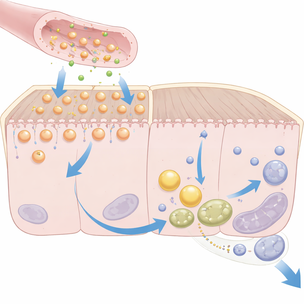
Protein processing in the early segment
In the early part of the tubule (called S1), the probes first appeared just under the brush border, then in early endosomes, and finally in small, highly acidified lysosomes clustered beneath larger vacuoles. There, protein degradation actually occurred. The researchers saw lysosomes repeatedly docking with and detaching from late endosomes, suggesting a busy hand-off zone for protein cargo. When they acutely neutralized lysosomal acidity with the drug hydroxychloroquine, protein uptake was strongly impaired, the key protein receptor megalin was misrouted away from the cell surface, and the normal dance between endosomes and lysosomes was frozen into large, fused structures. As a result, more protein leaked into the urine, mimicking features of kidney disease.
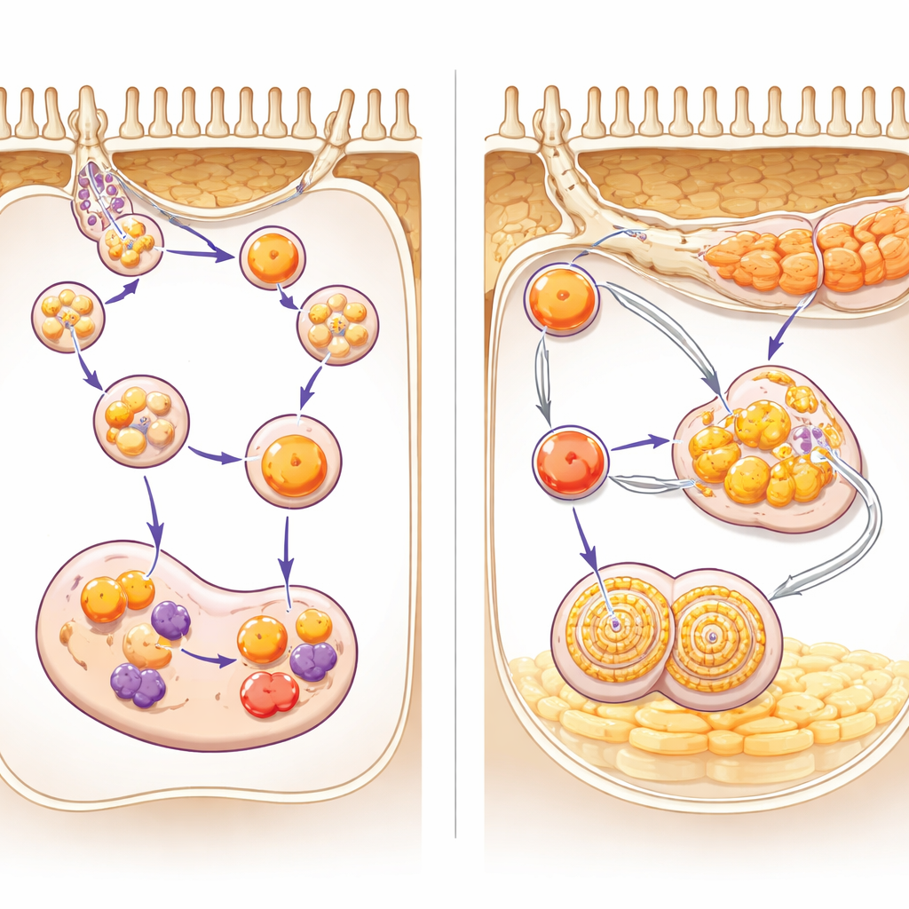
Fat handling in the downstream segment
Downstream, in the S2 segment, lysosomes told a different story. Here, they were larger, highly mobile, and rich in a lipid-digesting enzyme called lysosomal acid lipase. Imaging and electron microscopy showed fat-filled droplets clustered near mitochondria at the base of these cells. Lysosomes repeatedly traveled from the apical side down to this basal region, contacted lipid droplets, and sometimes appeared to wrap around and drag them through the cell. Over time, droplets were converted into multilamellar bodies — layered lipid-rich structures — that could be released into the tubular lumen. Blocking lysosomal lipase activity caused fats to build up near lysosomes, while chemically alkalinizing lysosomes redirected them away from the basal region and toward the lumen, promoting rapid lipid and multilamellar body secretion into urine.
What this means for kidney health
Together, these findings reveal that lysosomes in the proximal tubule are not generic trash cans but versatile, region-specific workhorses. In the early segment, they focus on recycling filtered blood proteins; in the later segment, they act as movers and grinders of fat, linking mitochondrial fuel use to lipid disposal. Disrupting their acidity — whether by drugs like hydroxychloroquine or by metabolic stress — scrambles these tasks, leading to protein loss and abnormal lipid handling. For a lay observer, the takeaway is that tiny shifts inside these microscopic structures can have big consequences for how kidneys manage nutrients and protect themselves from damage, offering fresh clues to how kidney diseases involving protein and fat imbalance may arise and how they might be treated.
Citation: Kaminska, M., Sakhi, I.B., Jankovic, N. et al. Heterogeneity in lysosomal dynamics and metabolic functions along the kidney proximal tubule. Nat Commun 17, 3677 (2026). https://doi.org/10.1038/s41467-026-70306-5
Keywords: kidney proximal tubule, lysosomes, protein reabsorption, lipid metabolism, hydroxychloroquine