Clear Sky Science · en
Parvalbumin-positive neurons in the medial septum participate in the formation of hippocampal-dependent spatial memory
Why remembering where things are can fail after a bad night
Have you ever noticed that after a sleepless night you misplace objects or struggle to remember where you left things? This study in mice looks inside the brain to find out why poor sleep makes it harder to remember where objects are, focusing on a small group of cells that link a deep memory region with a key control hub.
A small hub that talks to the brain’s map center
Our sense of place depends heavily on the hippocampus, a curved structure deep in the brain that builds an internal map of our surroundings. Within this map, special “place cells” fire when we are in specific locations, helping us track where we and nearby objects are. Another region, the medial septum, sends powerful control signals to the hippocampus and helps set its rhythmic activity. In this work, the authors zoomed in on a subset of medial septum cells that carry a protein called parvalbumin and release the calming chemical GABA. Earlier studies showed these cells influence brain rhythms linked to navigation, but it was unclear whether they directly shape memories that depend on the hippocampus.
Sleep loss, object-location memory, and brain rhythms
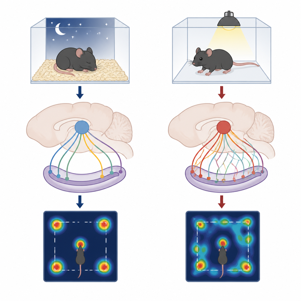
To mimic a rough night, the researchers gently kept male mice awake for five hours using a slow rotating bar that disturbed sleep without causing strong stress or anxiety. Afterward, the animals performed an object-place recognition task in a box containing two identical objects. First, both objects stayed in fixed corners while the mice explored and formed a memory; later, one object was moved to a new corner. Well-rested mice naturally spent more time investigating the moved object, indicating they noticed the change. Sleep-deprived mice explored just as much overall and moved normally, but their preference for the moved object dropped, showing that their object-location memory was impaired. At the same time, electrical recordings revealed that sleep loss reduced the strength of theta waves in the hippocampus and weakened the coordination between the medial septum and hippocampus during memory encoding and testing.
Neurons that fire for objects and guide the brain’s map
Using a combination of fine electrodes and light-based control tools, the team recorded activity from identified parvalbumin neurons in the medial septum along with hippocampal place cells while mice explored the objects. These septal neurons fired more strongly when mice were near objects, especially during the first phase when the memory was being formed, and different subgroups responded to each object. Their responses were largely independent of how fast the animal was moving, suggesting that they specifically carried information about objects rather than simple running speed. When many of these neurons were considered together, their joint activity could reliably distinguish which object the mouse was exploring. After sleep deprivation, however, their responsiveness to objects and their ability to distinguish between object locations dropped, particularly when one object had been moved.
How sleep loss scrambles the internal map
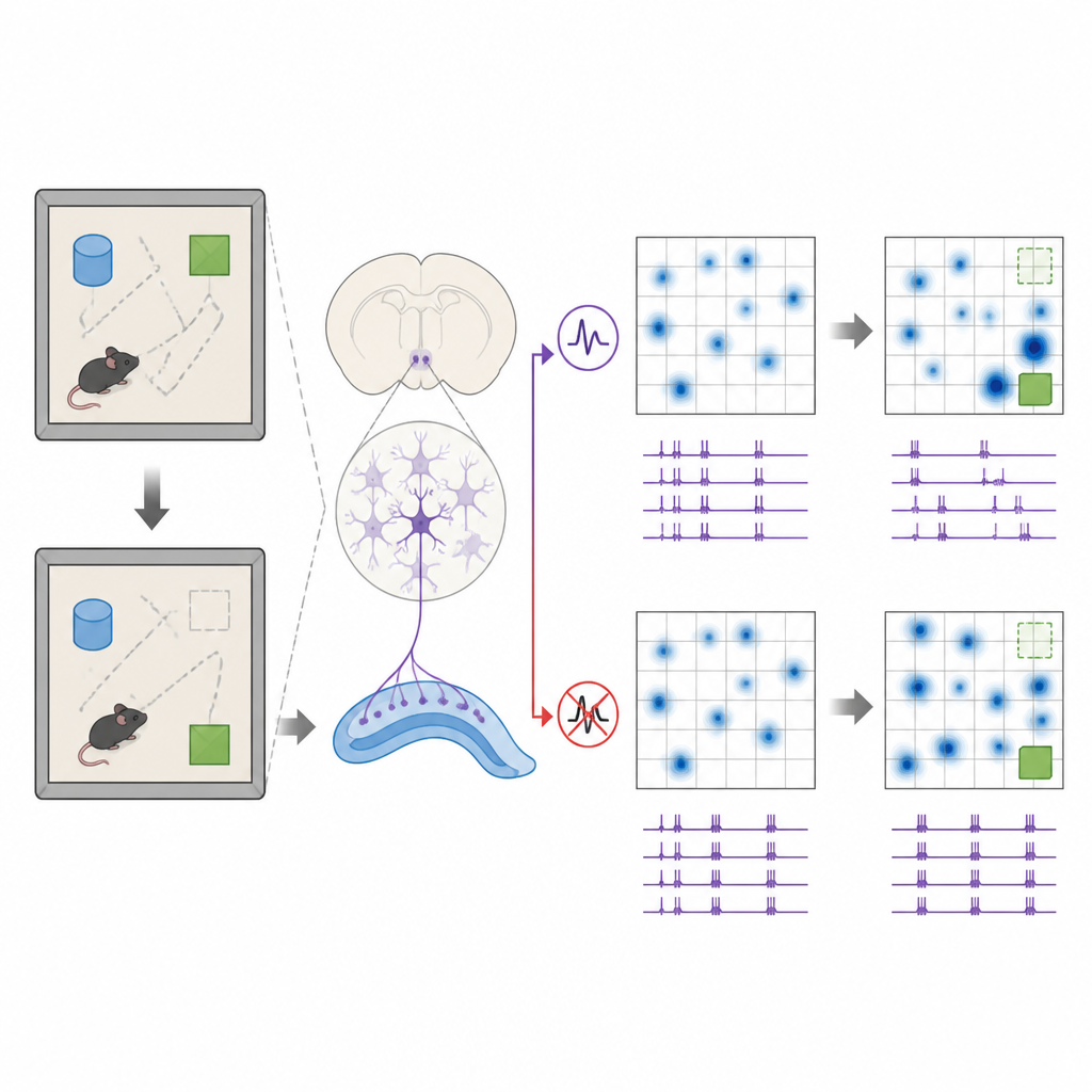
The authors then examined how place cells in the hippocampus updated their firing patterns when an object changed location. In rested animals, many place cells shifted their preferred firing spots toward the moved object, effectively updating the brain’s map to mark the new location. These shifts were biased in the direction of the moved object and were accompanied by a relatively flexible pattern of cooperation among place cells. After sleep loss, place cells were still present and active, but their firing fields shifted in a more random way, with fewer cells moving closer to the new object. At the same time, pairs of place cells became more tightly locked together in their activity, forming a more rigid network that appeared less able to reorganize when the environment changed.
Switching the pathway back on restores memory
To test cause and effect, the researchers used optogenetics to activate or silence parvalbumin neurons and their projections to the hippocampus during specific phases of the task. Briefly boosting their activity during the initial learning phase restored normal theta rhythms, loosened overly tight coupling among place cells, and brought back the tendency of place fields to shift toward the moved object, even after sleep deprivation. Behaviorally, this selective activation rescued the mice’s preference for the relocated object. In contrast, inhibiting these neurons or their direct pathway to the hippocampus disrupted object-place recognition, even without prior sleep loss, and activating them only during rest or retrieval phases had little benefit.
What this tells us about memory and poor sleep
For a layperson, the key message is that a small group of timing cells in the medial septum helps the hippocampus update its internal map when objects move, and that sleep loss blunts this fine control. When these cells cannot respond smartly to objects, the place cells that form our mental map become too rigid and fail to mark new locations accurately. By artificially turning this pathway back on at the right moment, the researchers could restore both the brain signals and the behavior in sleep-deprived mice. The findings suggest that the quality of communication between brain regions, rather than simple tiredness, underlies some of the memory slips we notice after a bad night’s sleep.
Citation: Zheng, Y., Tong, J., Xing, Y. et al. Parvalbumin-positive neurons in the medial septum participate in the formation of hippocampal-dependent spatial memory. Nat Commun 17, 4259 (2026). https://doi.org/10.1038/s41467-026-70268-8
Keywords: sleep deprivation, spatial memory, hippocampus, medial septum, place cells