Clear Sky Science · en
Assessing the micromechanical architecture of joint tissues with speckle rheological microscopy
Why joint softness and stiffness matter
Every step you take depends on a delicate balance between squishiness and springiness inside your knees and other joints. When this balance fails, as in osteoarthritis, joints become painful and stiff long before the damage is obvious on an X-ray. This study introduces a light-based microscope technique that can "feel" the inner mechanical landscape of joint tissues without touching or harming them, potentially revealing early warning signs of damage and guiding better treatments.
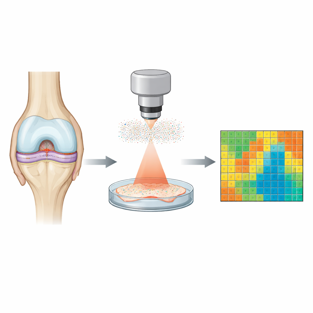
A new way to feel with light
The researchers developed Speckle Rheological Microscopy, or SHEAR, which measures how joint tissues resist and dissipate mechanical forces using only light. Instead of pressing or stretching samples, they shine a gentle laser on intact tissue and record a shimmering pattern called speckle. Tiny natural jitters of molecules and fibers inside the tissue—Brownian motion—make this pattern flicker over time. By analyzing how quickly and how strongly the speckle changes at each point, the system infers how firm (elastic) or fluid-like (viscous) the tissue is, and how this behavior changes across a range of time scales.
Seeing hidden patterns in healthy joints
To test SHEAR, the team examined cartilage, meniscus, tendon, ligament, and joint fluid from healthy swine knees. They created detailed maps showing overall firmness, how much energy is stored like a spring, and how much is lost like a shock absorber. A key index, called the dissipative fraction, highlights how fluid-like different regions are. These maps revealed fine structural patterns—such as beehive-like zones around cartilage cells or striping in ligaments—that were invisible in simple stiffness maps but matched microscopic anatomy. When they compared average values from SHEAR with those from a standard mechanical testing machine, the agreement was strong, confirming that this contact-free optical method can faithfully capture tissue mechanics.
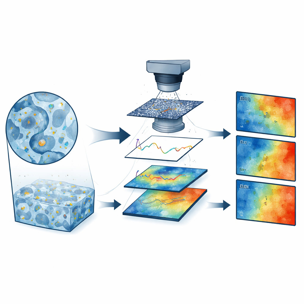
Tracking tiny injuries and repair
The technique was then used to examine small, laser-made defects in swine cartilage that had been allowed to heal for several months. Without cutting or isolating the cartilage from underlying bone, SHEAR could pick out each tiny lesion and distinguish a stiffer outer shell from a softer, more fluid-like core. The outer shell appeared mechanically reinforced, likely by fibrous repair tissue, while the center seemed under-built and more water-rich. These nuanced mechanical fingerprints were more sensitive than standard histology alone, suggesting that SHEAR could detect early or partial repair failures in ways that bulk tests and routine microscopy might miss.
Reading the mechanical fingerprint of diseased cartilage
The most striking results came from human knee cartilage removed during replacement surgery for advanced joint disease. In these samples, SHEAR maps showed dramatic shifts in both firmness and fluid-like behavior in regions with depleted cartilage molecules and disrupted collagen fibers. Superficial layers that looked worn and thinned under the microscope appeared softer and more dissipative, consistent with increased free water flow through a damaged matrix. Deeper, better-preserved regions remained relatively springy. Across multiple cases, areas poor in key cartilage molecules became mechanically weaker and more fluid-like, and these traits changed in distinctive ways across different probing frequencies. This frequency-dependent view captured how diseased tissue might respond differently to slow everyday movements versus faster or more forceful loads.
What this could mean for joint health
Taken together, the work shows that SHEAR can map the hidden mechanical architecture of joint tissues in fine detail, separating the contributions of solid fibers and trapped fluid. Because it is non-destructive, non-contact, and compatible with intact specimens of many shapes, it could become a powerful research tool for tracking how injuries heal, how arthritis progresses, and how engineered tissues mature. With further development into small probes, the same principle might eventually be used during arthroscopic surgery to provide a mechanical "heat map" of joint surfaces, helping clinicians spot early softening, guide treatment, and evaluate the success of repairs before damage becomes irreversible.
Citation: Leartprapun, N., Guastaldi, F.P.S., Randolph, M.A. et al. Assessing the micromechanical architecture of joint tissues with speckle rheological microscopy. Nat Commun 17, 3546 (2026). https://doi.org/10.1038/s41467-026-70248-y
Keywords: osteoarthritis, cartilage mechanics, optical microrheology, joint tissue imaging, degenerative joint disease