Clear Sky Science · en
Altering the carbohydrate-binding specificity of the legume lectin FRIL through structure-guided engineering
How Bean Proteins May Help Fight Viruses
Many viruses latch onto our cells by recognizing specific sugar patterns on the cell surface. If scientists can control how “sugar-sensing” proteins behave, they may be able to block infections or create better diagnostic tools. This study focuses on FRIL, a sugar-binding protein from the edible hyacinth bean, and shows how researchers reprogrammed it so that it changes which sugar structures it prefers to grab—potentially expanding its use as a broad antiviral and biomedical tool. 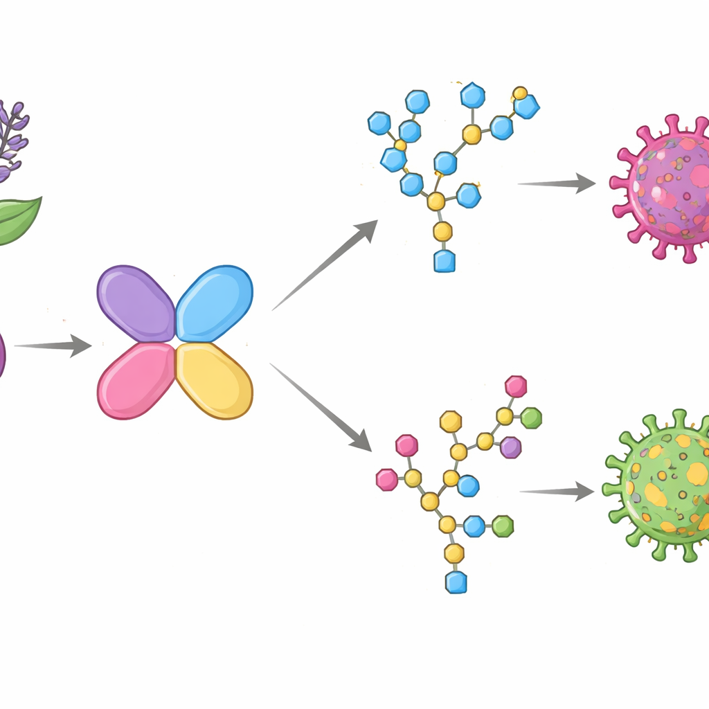
A Versatile Sugar-Detecting Bean Protein
FRIL belongs to a large family of plant proteins called legume lectins, which naturally bind to specific sugar decorations on proteins and cell surfaces. Unlike many related lectins that mainly prefer simple, tree-like mannose sugars, FRIL stands out because it favors more elaborate “complex” N-glycans—branched sugar chains that often carry extra building blocks such as fucose and galactose. FRIL has already been shown to preserve stem cells in culture, slow the growth of new blood vessels in tumors, and block viruses including influenza and SARS-CoV-2. These properties make it an attractive candidate for engineering, but reproducing the active bean protein in the lab had long been difficult.
Turning an Inactive Lab Product Into a Working Tool
When the team produced FRIL in mammalian cells, the protein folded correctly into its four-part structure but showed almost no sugar-binding activity. Using cryo-electron microscopy, they discovered why: FRIL carried its own sugar chain on a short linker region, and that chain folded back into the binding pocket like a built-in safety plug, blocking access to outside sugars. By removing these sugars with an enzyme that cuts N-glycans, they “unplugged” the pocket and restored full activity. The same trick worked on a related lectin precursor called proConA, suggesting a general strategy for producing active, engineered lectins from plants in standard lab systems.
Finding the Tiny Region That Sets Sugar Preference
With active FRIL in hand, the researchers solved high-resolution structures of the protein bound to two different sugars: a small piece of a complex N-glycan and a high-mannose chain. This allowed them to map which amino acids touched each part of the sugars. A few positions in a flexible loop, called loop B, stood out. In complex glycans, FRIL used two neighboring residues in this loop to form snug contacts with the extra fucose, galactose, and N-acetylglucosamine units. When the team mutated these two residues to smaller ones, FRIL did not lose activity—it lost its preference. The altered proteins now bound strongly to both complex and high-mannose chains, as confirmed by sugar arrays and binding tests with carefully glycosylated influenza proteins. 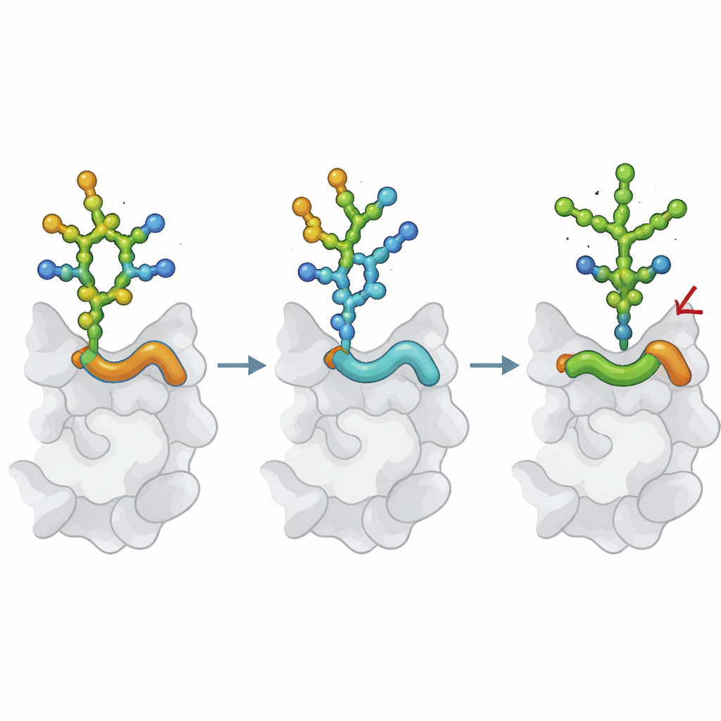
Rewiring FRIL to Prefer Virus-Like Mannose Patches
Guided by structural comparisons with Concanavalin A, a classic lectin that naturally prefers high-mannose sugars, the authors redesigned loop B more extensively and also changed a nearby loop C residue. This “FRIL-OMS” mutant shifted its sugar taste almost completely: it now favored high-mannose N-glycans and largely ignored the complex ones that wild-type FRIL prefers. New cryo-EM structures showed how the reshaped loop closed off the path used by complex glycans, while opening a new pocket that cradled an extra mannose branch. In laboratory virus tests, FRIL-OMS excelled at recognizing and neutralizing influenza strains whose surface proteins are heavily decorated with dense mannose patches, a hallmark of some circulating viruses.
Why This Matters for Future Medicine
This work shows that a few precise changes in a small surface loop can switch a lectin’s sugar preference from one type of N-glycan to another, while preserving its basic structure and safety profile. In practical terms, that means scientists can begin to “dial in” the sugar targets they want—whether to capture certain viruses, probe cancer-linked sugar patterns, or design new diagnostic arrays. FRIL and its engineered cousin FRIL-OMS illustrate how plant lectins can be converted into customizable, sugar-specific tools that may one day help detect disease earlier or block infection by locking onto the very sugar coats that viruses depend on.
Citation: Liu, YM., Nguyen, H.T.V., Chen, X. et al. Altering the carbohydrate-binding specificity of the legume lectin FRIL through structure-guided engineering. Nat Commun 17, 3528 (2026). https://doi.org/10.1038/s41467-026-70188-7
Keywords: lectins, glycans, protein engineering, antiviral, influenza