Clear Sky Science · en
Visualizing molecular diffusion direction and processes in the solid state via dichromatic fluorescent cocrystalization transformation
Watching Molecules Move in a Solid
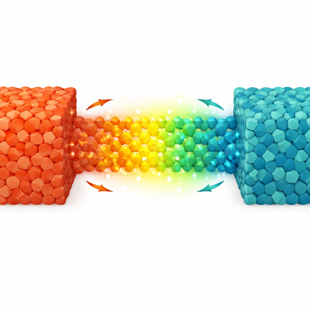
We tend to think of solids as rigid and unmoving, but at the molecular level they can be surprisingly dynamic. This study shows a way to actually “see” how molecules migrate through a solid, using color-changing crystals as a built‑in camera. Beyond the sheer visual appeal of glowing yellow and green crystals, the work matters because it offers a new tool for checking drug purity and tracking important chemical reactions in real time, without complex instruments.
Why Motion in Solids Matters
Even in a solid, molecules can twist, vibrate, and slowly drift past one another. These quiet motions underlie how materials change phase, stay stable over time, or respond to light and heat. Yet they are hard to study because the molecules are packed tightly together and cannot be watched directly. Traditional optical methods usually only tell us that something has changed, not how fast it changed, in which direction molecules moved, or what paths they followed. Researchers have been searching for simple, sensitive ways to track this hidden traffic inside crystals.
Building Color-Switching Crystals
The team tackled this problem by designing a pair of organic molecules that act like partners: one donates electrons and the other accepts them. When these two meet in the right arrangement inside a crystal, they share charge, which alters how they absorb and emit light. Using 6‑methoxy‑2‑acetylnaphthalene (a small drug‑like molecule, and an impurity related to the pain reliever naproxen) and a highly electron‑hungry compound called tetracyanobenzene, they created two distinct mixed crystals. One contains equal amounts of the two partners and glows yellow; the other has twice as much acceptor and glows green. The different colors arise from how tightly the molecules stack and how far apart the donor and acceptor columns sit within the crystal lattice.
Following Diffusion by Color
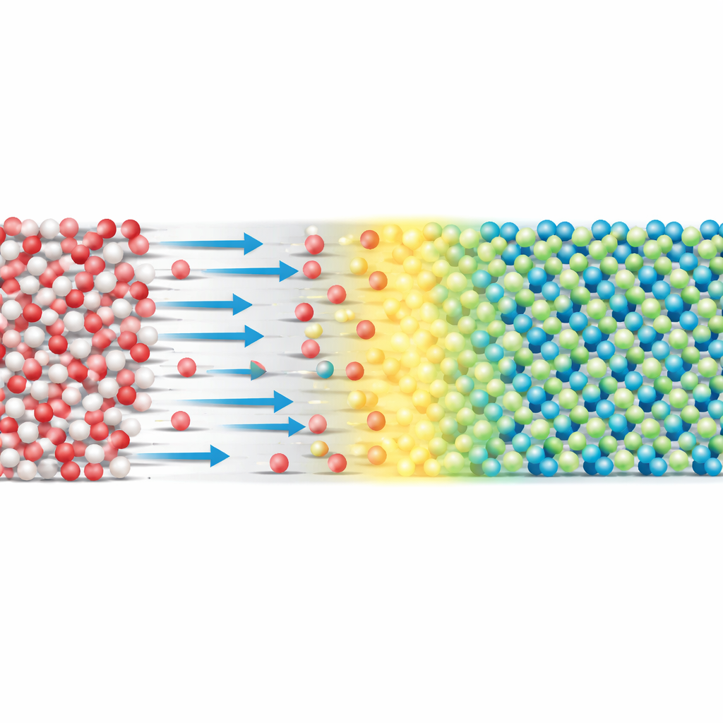
Because these two crystal types can convert into one another, the solid effectively acts as a color‑coded map of where and how the molecules have traveled. When the researchers simply pressed powders of the two components together, nothing seems to happen at first. Over minutes to hours, however, the contact region lights up. Yellow emission appears where molecules first meet and form the 1:1 crystal, and then, in regions rich in the acceptor, the color gradually shifts to green as the 1:2 crystal grows. Careful experiments in flat cuvettes showed a striking one‑way flow: the donor molecules diffuse deep into the acceptor region much faster than the reverse. This produces a moving front in which the interface glows yellow while the interior turns green, directly encoding both direction and speed of molecular diffusion in the pattern of colors.
From Glowing Crystals to Drug Quality Control
The same color‑sensitive behavior turns out to be very useful for pharmaceutical analysis. Naproxen, a common anti‑inflammatory drug, is known to carry the donor molecule as a key impurity at low levels. Unlike the impurity, naproxen itself barely interacts with the acceptor and does not produce strong charge‑transfer fluorescence. By grinding drug samples with the acceptor in different proportions, the authors could “light up” even 0.1% impurity content: first as yellow and then as green fluorescence, depending on how much acceptor was present. Related molecules with only slight structural changes did not trigger comparable color shifts, revealing a high chemical selectivity that helps avoid false positives.
Watching a Reaction as It Happens
The researchers pushed the method further by modifying naproxen to make a family of simple esters, which react with each other in a process called transesterification. Some of these esters form strong, yellow‑emitting charge‑transfer crystals with the acceptor, while others hardly respond. By exposing a solid mixture of esters and acceptor to ammonia vapor, they drove a transesterification reaction that slowly produced the “bright” ester. As it formed, the powder transitioned from faint blue emission to intense yellow glow, providing a direct visual readout of reaction progress in the solid state without dissolving the material or adding dyes.
What This Means in Simple Terms
In essence, this work turns a pair of small organic molecules into a built‑in sensor for motion and change inside solids. The yellow and green crystals act like traffic lights that show where molecules have moved, how fast they went, and what new structures they formed. By cleverly choosing donor molecules related to real medicines, the authors demonstrate that this colorful signal can flag tiny amounts of impurities and track useful chemical reactions as they unfold. The approach offers a vivid, accessible window into the normally invisible world of solid‑state molecular motion, with practical benefits for making safer drugs and better‑controlled materials.
Citation: Zheng, J., Zhu, X., Wang, W. et al. Visualizing molecular diffusion direction and processes in the solid state via dichromatic fluorescent cocrystalization transformation. Nat Commun 17, 3295 (2026). https://doi.org/10.1038/s41467-026-70152-5
Keywords: solid-state molecular motion, charge-transfer cocrystals, fluorescent sensing, naproxen impurity detection, molecular diffusion visualization