Clear Sky Science · en
Engineering NIR-II carbon dots through aniline extension with graphene and nitrogen enrichment for hepatobiliary theranostics
Seeing Deeper Inside the Liver
Surgeons and doctors increasingly rely on glowing dyes to see hidden structures inside the body, especially during delicate operations on the liver and gallbladder. But today’s dyes do not shine well through thick tissue and can miss tiny leaks or early signs of disease. This study introduces a new class of tiny glowing particles, called carbon dots, that can both light up the bile ducts in unprecedented detail and help treat liver scarring, offering a glimpse of future tools for safer surgery and earlier therapy.
Tiny Lights Built from Everyday Rings
The researchers set out to redesign carbon dots so that they emit light in the so‑called “second near‑infrared window,” a range of wavelengths that travels more deeply and clearly through the body than visible light. They started from simple ring‑shaped molecules related to aniline, a common building block in chemistry, and linked them into increasingly extended frameworks. During a one‑pot heating process with selenourea, these frameworks carbonized into three kinds of carbon dots (CDs‑1, CDs‑2, and CDs‑3), whose glow could be tuned from visible blue‑green all the way to the deep near‑infrared. Electron microscopy and spectroscopy showed that, as the dots grew, they developed more ordered, graphene‑like patches and a higher content of specific nitrogen forms in their structure.
How Structure Turns Color into Deep Infrared
To understand why the glow shifted to longer wavelengths, the team combined detailed measurements with computer calculations. By gradually adding aniline units, they strengthened the separation between regions that donate electrons and those that accept them inside each precursor molecule. This increased the molecular dipole moment and made it easier for electrons to move across the structure, lowering the energy gap between electronic states. As the dots carbonized, graphene‑like domains expanded and a particular nitrogen type, called pyrrolic nitrogen, accumulated. Modeling showed that these features further widened the pathway for electrons and narrowed the energy gap to values far below those of ordinary dyes, pushing the emission into the NIR‑II region. In CDs‑3, this produced strong emission peaks around 1080 and 1265 nanometers, a regime rarely reached by intrinsic carbon materials.
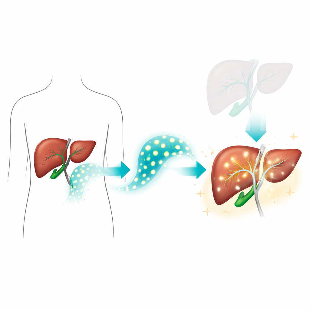
Lighting Up Bile Ducts and Hidden Leaks
With these properties in hand, the scientists tested whether CDs‑3 could improve imaging of the gallbladder and bile ducts, structures that are easily injured during keyhole surgery. In human gallbladder tissue models and animal studies, the dots were secreted into bile and produced bright NIR‑II signals that remained visible through up to 15 millimeters of overlying tissue—far beyond the roughly 2 millimeters achieved by the standard hospital dye indocyanine green. Using different optical filters, they could clearly outline normal bile ducts, pinpoint strictures created by ligatures, and detect small leaks where bile escaped into surrounding tissue. Signal‑to‑noise ratios and image sharpness were high enough to resolve sub‑millimeter features, suggesting that such probes could give surgeons a real‑time map of the biliary tree with far greater confidence.
From Diagnosis to Treatment of Liver Scarring
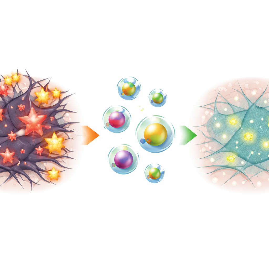
Because liver disease is often driven by excessive reactive oxygen species—highly reactive molecules that damage cells and promote scarring—the team also explored a therapeutic angle. The carbonization recipe, which includes selenium‑containing selenourea and electron‑rich aniline structures, naturally endowed CDs‑3 with strong antioxidant properties. To steer the dots more selectively to scar‑forming liver cells, they coated them with a biodegradable polymer bearing a short peptide that homes to hepatic stellate cells. The resulting composite, called CDs‑3@pPB, formed roughly 100‑nanometer particles that are well suited to accumulate in the liver. These particles remained dark when clustered, but in oxidant‑rich environments such as fibrotic tissue, they broke apart, brightened, and released active carbon dots—turning high oxidative stress into both a trigger for treatment and a stronger imaging signal.
Slowing and Revealing Liver Fibrosis
In cell cultures, CDs‑3@pPB quenched several types of reactive oxygen species and protected liver cells from oxidative damage. In activated stellate cells, it lowered key markers of scarring and reduced cell proliferation more effectively than a reference liver drug, silymarin. In mouse models where liver fibrosis was induced by a toxic chemical, treated animals showed smoother liver surfaces, improved blood enzyme levels, less collagen buildup, and fewer activated stellate cells compared with untreated controls. Importantly, NIR‑II imaging with CDs‑3@pPB revealed stronger signals in damaged livers than in healthy ones, tracking cell death and scar formation without the need for invasive biopsies, while safety studies showed minimal side effects and good clearance over time.
What This Could Mean for Patients
Taken together, this work demonstrates that carefully engineered carbon dots can do double duty as deep‑penetrating flashlights and active medicines for the liver and bile ducts. By tailoring molecular building blocks and carbonization conditions, the authors created NIR‑II probes whose color shift arises from their evolving internal structure, rather than from attached dyes. CDs‑3 enables clearer visualization of bile ducts and leaks during surgery, while its liver‑targeted cousin CDs‑3@pPB can both reveal and relieve liver fibrosis in preclinical models. Although further studies will be needed before human use, this approach sketches a path toward tiny, smart particles that help doctors see and heal liver disease in the same step.
Citation: Yang, L., Li, M., Peng, Y. et al. Engineering NIR-II carbon dots through aniline extension with graphene and nitrogen enrichment for hepatobiliary theranostics. Nat Commun 17, 3336 (2026). https://doi.org/10.1038/s41467-026-70150-7
Keywords: near-infrared imaging, carbon dots, bile duct surgery, liver fibrosis, nanotheranostics