Clear Sky Science · en
Structural dynamics and immunogenicity of the recombinant and outer membrane vesicle-embedded Meningococcal antigen NadA
Why this study matters for future vaccines
Vaccines work best when they show our immune system disease targets in the same way these targets appear on real microbes. This study asks a deceptively simple question with big implications: does it matter if a key meningitis vaccine protein is given as a free-floating molecule, or displayed in a tiny bubble of bacterial membrane that mimics its natural home? The answer could change how we design safer and more powerful vaccines against tough bacterial infections.
The bacterial protein at the heart of protection
The work centers on NadA, a surface protein from Neisseria meningitidis, the bacterium that causes deadly meningitis B. NadA helps the microbe latch onto cells in our airways and is one of the main protective components of the licensed 4CMenB vaccine. In the current vaccine, NadA is used in a trimmed, soluble form that is easier to manufacture than the full membrane-anchored version. However, not all antibodies raised by this soluble version can recognize NadA as it appears on the real bacterium, raising concerns that subtle differences in shape might limit protection.
Probing shape and motion with molecular “stop-motion”
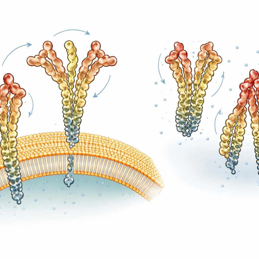
To uncover how NadA behaves in different environments, the researchers used hydrogen–deuterium exchange mass spectrometry, a technique that works like molecular stop-motion photography. Parts of the protein that are rigid and well-packed swap their hydrogen atoms slowly, while flexible or exposed regions swap them quickly. By monitoring these exchanges along the entire length of NadA, the team could infer where the protein forms stable coiled structures, where it bends, and where it behaves like a loose tail. They combined this with cryo–electron microscopy snapshots to confirm that the soluble NadA forms a long, flexible, three-part structure with a compact head, a rod-like stalk, and an unstructured tail.
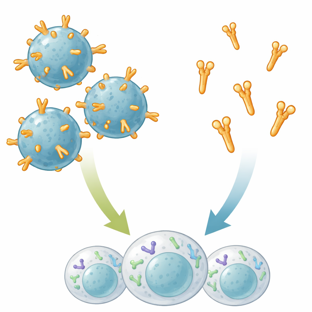
Native membrane bubbles change how NadA moves
The scientists then examined NadA in its native-like setting: outer membrane vesicles (OMVs). These are tiny spheres naturally shed by bacteria that preserve the same outer membrane and proteins found on the cell surface. When NadA was embedded in OMVs, several parts of the protein became more rigid compared with the soluble form, especially near the membrane anchor and along sections of the stalk and head. At the same time, the data revealed two coexisting shapes of the NadA trimer: a more tightly packed form and a more open form, akin to a breathing motion. In OMVs, the “open” version of the head region was more common than in the soluble protein, suggesting that the membrane anchor transmits mechanical constraints along the stalk that encourage the trimer to partially open and expose more surface area.
A stronger and more efficient immune punch
To see whether these structural shifts matter for protection, mice were immunized either with the soluble NadA protein or with OMVs carrying full-length NadA on their surface. Both approaches induced similar amounts of NadA-specific antibodies. However, when the researchers tested how well these antibodies could kill live meningococcal bacteria, the difference was striking: sera from OMV–NadA–immunized mice showed bactericidal activity more than an order of magnitude higher than sera from mice that received soluble NadA, even though the OMVs delivered much less NadA by weight. This suggests that presenting NadA in its natural membrane setting not only displays more relevant shapes and binding sites, but also may cluster the protein in a way that better activates B cells.
What this means for next-generation vaccines
In accessible terms, the study shows that “how” a vaccine antigen is displayed can be as important as “what” it is. When NadA is anchored in membrane bubbles that mimic the bacterial surface, it adopts slightly different, more dynamic shapes that appear to reveal key targets for protective antibodies. Those antibodies are then better at recognizing and killing real meningococcal cells. The findings support using native-like platforms such as OMVs or nanoparticles to present bacterial proteins in future vaccines, and suggest that deliberately encouraging an “open” form of trimeric antigens could be a promising strategy to boost vaccine effectiveness.
Citation: Calvaresi, V., Dello Iacono, L., Borghi, S. et al. Structural dynamics and immunogenicity of the recombinant and outer membrane vesicle-embedded Meningococcal antigen NadA. Nat Commun 17, 3777 (2026). https://doi.org/10.1038/s41467-026-70059-1
Keywords: meningococcal vaccines, outer membrane vesicles, NadA antigen, protein conformation, structural vaccinology