Clear Sky Science · en
Reversible surface modifications of functional proteins for accelerated cytosolic delivery via cell-penetrating peptide clusters
Bringing Working Proteins Inside Living Cells
Many modern medical and biological ideas depend on getting fully formed proteins into the interior of living cells, where they can act as tools, sensors, or even medicines. Yet proteins are large, fragile molecules that usually bounce off the cell’s outer membrane. This study introduces a simple, reversible way to “repackage” a wide variety of proteins so they can slip into the cell’s watery interior—without losing their function—and even works in the more stubborn cells of plants.
Why Getting Proteins Inside Cells Is So Hard
Cells are guarded by membranes that keep most large molecules out. Over the years, scientists have tried many tricks to smuggle proteins across this barrier. One of the most promising uses short chains of amino acids called cell‑penetrating peptides, which carry other molecules into cells. A powerful version of this idea uses clusters of a positively charged peptide known as Tat to drag in antibodies. However, this approach only works well for a narrow set of proteins and often requires high, sometimes toxic, doses. Proteins differ greatly in size and overall charge, and many simply do not interact properly with Tat clusters, so they remain stranded outside or trapped in internal bubbles rather than reaching the cell interior.
Giving Proteins a Temporary “Velcro Patch”
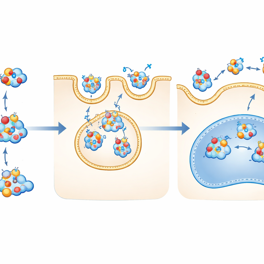
The researchers discovered that many difficult‑to‑deliver proteins can be helped by giving them a small, reversible “anionic patch” on their surface. This patch is a short, negatively charged peptide that can be clipped onto exposed sulfur‑containing sites on a protein by a chemical link called a disulfide bond. The negative patch strongly attracts the positively charged Tat3 peptide clusters, forming mixed complexes that cells eagerly take up. Once inside the cell’s reducing environment, the disulfide bond is naturally broken, the patch falls off, and the original protein is released in its native form. By carefully testing a series of patch designs, the team identified one, called E4D3, that balances strong attraction to Tat3 with efficient release inside cells.
Delivering Many Proteins, From Enzymes to Antibodies
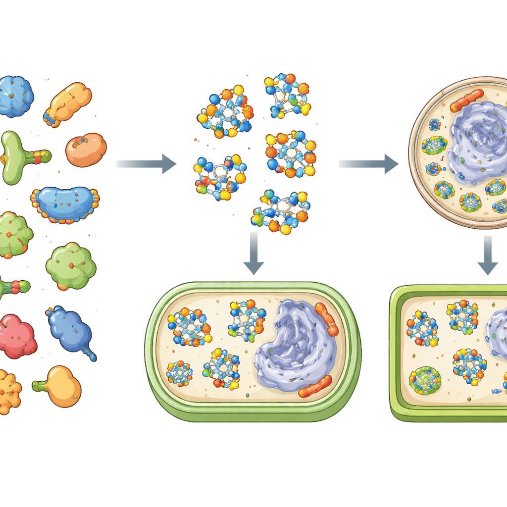
Using this strategy, the authors delivered a broad range of proteins into human cells at low micromolar concentrations. These included very small targeting peptides, fluorescent marker proteins, enzymes that cut RNA, large antibodies, and giant enzyme complexes weighing up to 430 kilodaltons. Proteins with a wide spread of overall electrical charges—from strongly acidic to strongly basic—could be brought into the cytosol, the fluid interior where most cellular chemistry happens. Importantly, the delivered proteins remained active: an RNA‑cutting enzyme selectively killed cells once inside, other enzymes carried out color‑changing reactions inside their new hosts, and a peptide that binds the cell’s internal scaffolding lit up the actin network in living cells.
How the Proteins Get In and What Happens Next
To understand the path into the cell, the team tracked fluorescently tagged proteins and used chemical blockers of different uptake routes. They found that the patched proteins bound to Tat3 through simple charge attraction, then entered cells mainly via macropinocytosis—a process where the cell membrane ruffles and engulfs nearby material into large pockets. Once inside, most of the protein–Tat3 complexes escaped from acidic compartments and spread through the cytosol and nucleus. The same method succeeded in plant leaves, which adds an extra obstacle in the form of a rigid cell wall, suggesting that the approach is robust across very different cell types.
Mapping Protein Networks Inside Living Cells
The authors also showcased a more advanced application: delivering a custom‑built protein probe that links a particular kind of “tagging” enzyme (an E2) to ubiquitin, a small modifier protein. This probe can latch onto and capture partner enzymes (E3 ligases) when activated by light, allowing researchers to identify them by mass spectrometry. Using the delivery method, they introduced this probe into living human cells and mapped dozens of E3 partners under growth‑factor stimulation, revealing a detailed interaction network under realistic, physiological conditions rather than in broken‑cell extracts.
What This Could Mean for Future Therapies and Tools
In plain terms, this work shows that adding a small, removable “charge handle” to a protein lets it hitch a ride with a peptide carrier into many kinds of cells, including hard‑to‑penetrate plant tissues. Because the handle falls off inside, the protein arrives in its original working form. This simple, mix‑and‑go chemistry could make it much easier to use tailor‑made proteins as research tools and, eventually, as treatments that act directly inside cells, expanding the toolbox for everything from basic cell biology to precision medicines.
Citation: Hua, X., Guo, Y., Li, P. et al. Reversible surface modifications of functional proteins for accelerated cytosolic delivery via cell-penetrating peptide clusters. Nat Commun 17, 3341 (2026). https://doi.org/10.1038/s41467-026-70054-6
Keywords: intracellular protein delivery, cell-penetrating peptides, protein engineering, macropinocytosis, ubiquitin signaling