Clear Sky Science · en
Cryogenic electron tomography reveals herpesvirus capsid assembly intermediates inside the cell nucleus
Why tiny virus shells matter to our health
Herpesviruses, including the germs behind chickenpox, shingles, cold sores, and several cancers, protect their genetic material inside a tough protein shell, or capsid. How these microscopic shells form and mature inside our cells helps determine whether infection succeeds or fails. This study uses cutting-edge cryogenic electron tomography—essentially 3D electron microscopy on flash-frozen cells—to watch herpesvirus shells assembling inside the cell nucleus, revealing hidden intermediate stages that could become new targets for antiviral drugs and vaccines. 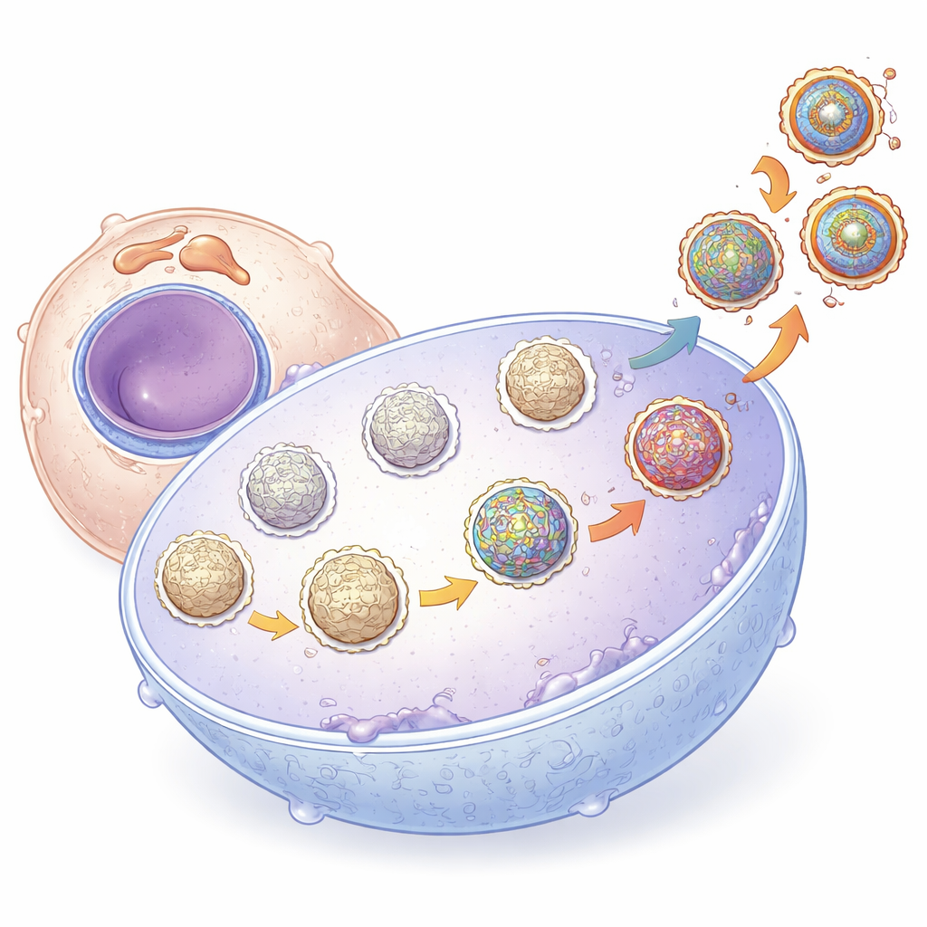
From chickenpox virus to a broader herpes family story
The researchers focused on varicella-zoster virus, the herpesvirus that causes chickenpox and shingles, but their findings are relevant to the entire herpes family. These viruses share a common architecture: a four-layered particle with a DNA core wrapped in a capsid, surrounded by a protein-rich tegument and a fatty envelope. The capsid is built in the host cell nucleus, where newly made shells first form around a temporary inner framework of scaffold proteins before being filled with viral DNA. Historically, scientists classified purified capsids into three types—A (apparently empty), B (containing scaffold), and C (packed with DNA)—but it has remained controversial whether A and B capsids are dead ends or true steps on the way to infectious particles.
Seeing virus assembly inside intact cells
To answer this, the team combined focused ion beam milling with cryogenic electron tomography. They first grew human skin cells on specially patterned grids, infected them with a fluorescently tagged virus, and rapidly froze them in a lifelike state. Using a focused ion beam, they shaved the cells down to thin slices suitable for electron imaging, then collected tilt series to reconstruct 3D volumes at near-atomic detail. These tomograms captured multiple stages of the viral life cycle in their natural setting: newly formed shells in the nucleus, capsids budding through the nuclear membrane, particles acquiring tegument and envelope in the cytoplasm, and fully formed virions poised at the cell surface.
Uncovering hidden stages in shell maturation
High-resolution reconstructions of more than a thousand capsids revealed that truly empty A-type shells are essentially absent in the nucleus. Instead, objects that once looked empty in thin 2D slices usually contain residual scaffold protein when viewed in 3D, suggesting that earlier work partly misclassified these structures. The authors applied advanced computational clustering to group capsids by their internal contents and outer decorations. They found a continuum: early shells were spherical and filled with dense scaffold; later ones became more angular, with less scaffold and eventually tightly packed DNA. Crucially, they correlated these interior changes with the appearance of a specialized five-part protein assembly at each capsid corner called the capsid vertex-specific component, or CVSC. 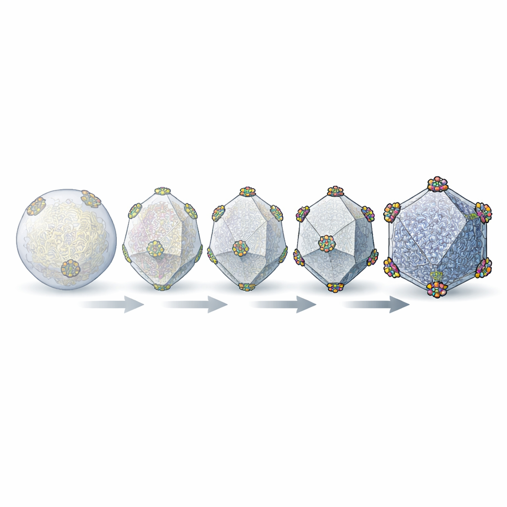
A molecular “timestamp” on the virus shell
The CVSC acts like a locking and reinforcing clamp at the vertices of the capsid. By focusing on each corner in turn, the team counted how many of these complexes were present per capsid. Capsids still rich in scaffold carried only a few CVSC units. Intermediate shells with less scaffold had more CVSCs, while fully DNA-filled capsids had nearly complete coverage—up to the maximum of five complexes at each of the twelve vertices. The researchers also resolved, for the first time in cells, the specialized “portal” vertex through which DNA is pumped into the shell. In DNA-filled capsids this portal is capped and blocked, consistent with a locked, pressurized container, whereas in scaffold-containing intermediates the portal region remains open and lacks the full cap, indicating a shell still preparing for genome packaging.
What this means for fighting herpes infections
Putting these pieces together, the study proposes that capsid assembly is not a series of hard jumps from A to B to C, but a smooth progression in which scaffold levels fall and CVSC occupancy rises as the shell matures and becomes ready to accept and retain the viral genome. Rather than being useless byproducts, many structures previously lumped together as A- or B-capsids are now reinterpreted as true intermediates with the potential to become infectious particles. Because the CVSC both stabilizes the DNA-filled capsid and helps it leave the nucleus, its gradual acquisition effectively timestamps how far along each shell is in the assembly line. By revealing this in-cell choreography at near-atomic resolution, the work highlights specific molecular interactions—especially at the capsid vertices and portal—that could be disrupted by future drugs to destabilize the capsid, block genome packaging, or prevent mature particles from exiting the nucleus.
Citation: Oliver, S.L., Chen, M., Engel, L. et al. Cryogenic electron tomography reveals herpesvirus capsid assembly intermediates inside the cell nucleus. Nat Commun 17, 3197 (2026). https://doi.org/10.1038/s41467-026-69811-4
Keywords: herpesvirus capsid, cryo-electron tomography, virus assembly, varicella-zoster virus, capsid maturation