Clear Sky Science · en
Mechanochemical feedback between confinement and actin crosslinking drives the shape dynamics of liquid-like droplets
How Soft Droplets Help Cells Shape Their Skeleton
Inside our cells, many key molecules gather into tiny, liquid-like droplets that lack traditional membranes. This study shows that when such droplets trap growing actin filaments—the protein rods that give cells their shape—they do more than passively hold them. The droplets and filaments push and pull on each other, reorganizing into rings, discs, and rods that can dramatically change droplet shape. Understanding this hidden mechanical partnership sheds light on how cells move, divide, and sense their environment, and may clarify what goes wrong in diseases where cell shape and motion are disrupted.
Protein Droplets as Tiny Construction Sites
The authors focus on biomolecular condensates: soft, liquid-like clusters of proteins that behave like droplets. Many actin-binding proteins can phase-separate into such droplets and recruit actin, turning the droplets into miniature construction sites for the cell’s internal skeleton. In these crowded spaces, simple actin filaments can be transformed into complex networks that underlie structures such as cell edges, contractile rings, and stress fibers. But how the droplets’ own physical properties—like surface tension—and the binding behavior of actin crosslinkers shape these networks has been poorly understood.
Simulations Meet Test-Tube Experiments
To tackle this question, the team built an agent-based computer model and paired it with carefully controlled laboratory experiments. In the simulations, individual actin filaments grew inside a deformable, ellipsoid-shaped droplet. Proteins such as VASP or lamellipodin were represented as crosslinkers that can join filaments together, either as fixed four-armed units or as dynamic chains that assemble and break apart. The droplet’s surface tension resisted deformation, while the growing and bending filaments pushed back on the boundary. Parallel experiments recreated similar droplets containing purified actin and actin-binding proteins, allowing the researchers to directly compare predicted shapes with real microscopic images.
From Rings and Discs to Snapping Droplets
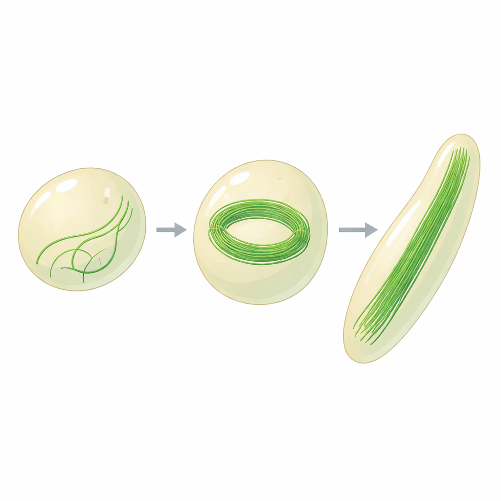
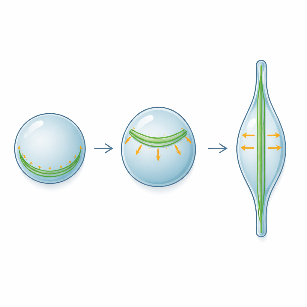
The combined approach revealed two main kinds of actin structures inside droplets: tightly bundled rings and more weakly bundled, disc-like arrangements. When the boundary was rigid, actin tended to form shells or rings that hugged the inner surface. Once the droplet was allowed to deform, those same filaments could instead collect into thick discs aligned with the direction in which the droplet stretched, minimizing their bending. Strikingly, the thickness of the actin bundle needed to deform a droplet increased with droplet diameter following a power-law rule, confirmed across simulations and experiments and for several different crosslinker types. The timing of shape change was also rich: droplets could temporarily stretch, relax toward a more spherical shape, and then “snap” into a more elongated form as filaments reorganized—behavior reminiscent of mechanical snap-through in everyday objects like bent plastic strips.
Filament Length, Crosslinkers, and Even No Crosslinkers at All
The study shows that filament length is a key control knob. Introducing capping proteins, which stop filaments from growing, shortened them and reduced droplet deformation both in silico and in vitro. Variants of crosslinkers that dynamically multimerize allowed filaments to rearrange more freely, often producing higher aspect-ratio droplets than rigid, tetrameric VASP. Surprisingly, the researchers also tested droplets that lacked any specific crosslinkers and found that confinement and droplet mechanics alone could bundle actin into discs and deform the droplet. Experiments with RGG protein condensates—which interact only weakly with actin—confirmed that simply packing growing filaments into a soft boundary is enough to generate bundles and rod-like droplet shapes.
Why This Matters for Cell Shape and Disease
Overall, the work establishes a general mechanochemical feedback loop: the droplet’s surface tension and viscosity set how easily it can deform, while actin growth and crosslinking determine how much bending energy is available to reshape it. Larger, tighter bundles exert stronger forces, and the number of filaments required to deform a droplet rises predictably with droplet size. These principles likely extend beyond the proteins studied here to many condensates that interface with the cytoskeleton, such as those at nerve terminals or cell adhesion sites. By showing that even simple physical rules can generate complex, dynamic shapes, the study offers a powerful framework for understanding how cells sculpt their interior architecture—and how subtle changes in protein interactions could tip that balance in disease.
Citation: Mansour, D., Jordan, D., Walker, C. et al. Mechanochemical feedback between confinement and actin crosslinking drives the shape dynamics of liquid-like droplets. Nat Commun 17, 3068 (2026). https://doi.org/10.1038/s41467-026-69803-4
Keywords: actin cytoskeleton, biomolecular condensates, phase separation, cell mechanics, protein droplets