Clear Sky Science · en
Cryo-EM structures of light chain fibrils from abdominal fat biopsies of multiple myeloma patients
Why this matters for patients and families
When people hear about multiple myeloma, they usually think of a cancer of the bone marrow. But for many patients, an invisible second threat is quietly building up in their organs: tiny protein fibers called amyloid. This study looks directly at those fibers, taken from living patients through a small fat biopsy, and reveals their atomic structure. Understanding how and why these fibers form helps explain why some myeloma patients become much sicker, and it may open the door to earlier diagnosis and more tailored treatments.
Two related diseases, one shared culprit
Multiple myeloma is a cancer of plasma cells, the immune cells that normally produce antibodies to fight infection. In many patients, these cancer cells overproduce small antibody fragments known as light chains, which circulate in the blood at very high levels. A related condition, called light chain amyloidosis, also involves these same proteins, but there the main problem is that light chains misfold and clump into long, rigid fibers that clog organs such as the heart and kidneys. Some people have both diseases at once, and they fare worse than patients with myeloma alone. Why the same type of protein behaves differently in these two settings has been a long-standing puzzle.
Looking inside fat tissue for hidden fibers
To tackle this question, the researchers used abdominal fat-pad biopsies, a routine and relatively gentle procedure already used to look for amyloid deposits. From fat samples of two patients with multiple myeloma and coexisting light chain amyloidosis, they isolated abundant protein fibers. Using cryo-electron microscopy, a technique that images flash-frozen samples with a beam of electrons, they reconstructed the three-dimensional shapes of these fibers at near-atomic detail. 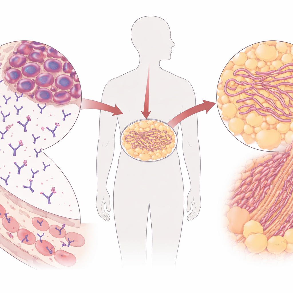
Three distinct fiber shapes from only two patients
The team discovered that light chains from these myeloma patients formed three different fiber architectures. In one patient, the same light chain sequence produced two polymorphs: one built from a single strand of protein winding into a helix, and another in which two such strands twisted together side by side. In the second patient, only one, thinner fiber type was observed. Despite these differences, all fibers showed a compact “core” made mainly from the variable part of the light chain—precisely the segment that normally helps antibodies recognize germs. The study also identified extra, cloud-like densities decorating the surfaces and at contact points between strands, hinting at additional molecules or subtle chemical changes that may help stabilize the structures.
Same building blocks, different ways to go wrong
To understand how these myeloma-derived fibers compare to those from light chain amyloidosis alone, the authors matched their structures to previously solved fibers from other patients. They found that some myeloma fibers resembled known amyloid structures but had notable differences: fewer rigid sheet-like segments, opposite twist directions, or missing structural elements seen in amyloidosis cases with many mutations. Computer modeling showed that in classic light chain amyloidosis, numerous mutations tend to destabilize the normal, soluble form of the light chain, making it easier for the protein to misfold and aggregate. In contrast, the light chains from the myeloma patients were much closer to their original “germline” sequences and remained relatively stable as individual molecules. 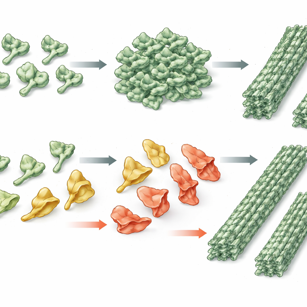
What this means for care and future research
In plain terms, this work shows that amyloid fibers in multiple myeloma patients can be built from light chains that are not dramatically misshapen on their own but are present at such high levels that they eventually stack into harmful deposits. At the same time, the final fibers are just as sturdy and difficult to dissolve as those seen in classic light chain amyloidosis. By revealing these structures directly from an easy-to-access fat biopsy, the study provides a molecular blueprint that could improve how doctors detect and classify amyloid in living patients. It also suggests that myeloma therapies that reduce light chain production early and aggressively might help prevent dangerous buildup, complementing future drugs designed to block misfolding or fiber growth.
Citation: Yao, Y., Yao, S., Xu, Y. et al. Cryo-EM structures of light chain fibrils from abdominal fat biopsies of multiple myeloma patients. Nat Commun 17, 3137 (2026). https://doi.org/10.1038/s41467-026-69784-4
Keywords: multiple myeloma, light chain amyloidosis, amyloid fibrils, cryo-EM, protein aggregation