Clear Sky Science · en
Sono-mechanical nanostructures-enabled sustained precise ultrasound brain stimulation
Listening to the Brain with Gentle Sound
Brain disorders like Parkinson’s disease are often treated with deep brain stimulation, which requires surgically implanted electrodes. This study explores a very different idea: using gentle ultrasound waves, guided by tiny engineered particles, to nudge specific brain cells without surgery or genetic modification. For a lay reader, the appeal is clear—if this approach can be made safe and precise in humans, it could offer a new, less invasive way to treat movement disorders and study how the brain works over long periods of time. 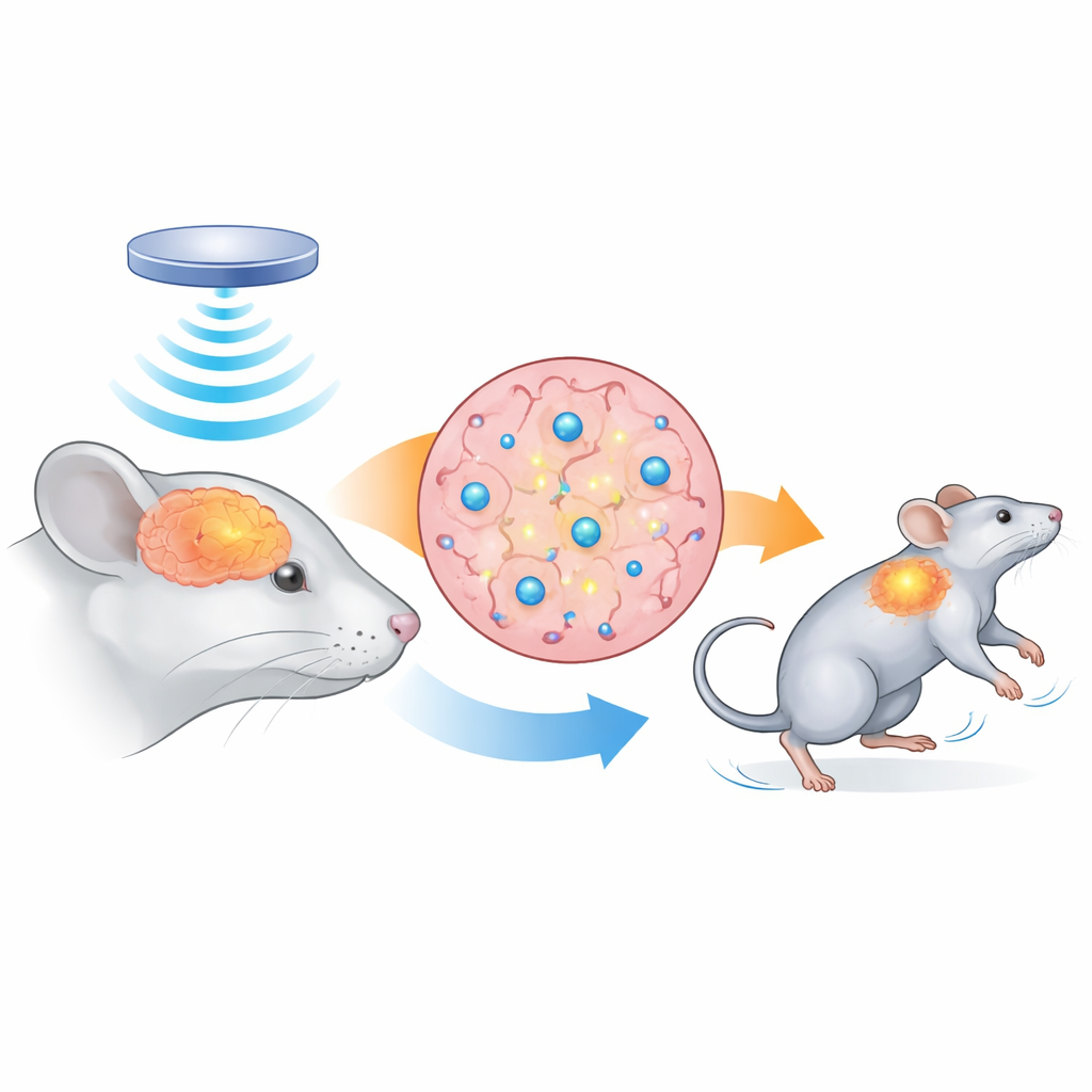
Tiny Echo Chambers Built for Sound
The researchers designed hollow silica nanostructures—essentially microscopic shells with a gas-filled core—that act like miniature echo chambers for ultrasound. Their rigid silica walls and gas interior make them vibrate strongly when hit by sound waves, concentrating mechanical energy at their surface. The team coated these shells with biocompatible polymers and iron, which helps them remain stable in the brain, disperse well in fluid, and be tracked by MRI and ultrasound imaging. Lab tests confirmed that these particles are uniform in size (about one-fifth of a micrometer wide), stable under repeated ultrasound, and non-toxic to cultured neurons.
Turning Sound into Nerve Activity
To see whether these hollow particles could help control brain cells, the team first worked with neurons grown in dishes. When they added the nanostructures and applied low-intensity ultrasound, calcium flooded into the neurons—a clear sign that the cells had fired. This effect depended on the hollow design: solid silica particles did not work. It also depended on special “mechanosensitive” channels in the cell membrane, which open when the membrane is pushed or stretched. When the researchers blocked these channels with a drug, the sound-plus-nanostructure effect largely disappeared and then returned when the drug was washed away. In short, the particles acted as amplifiers that turned mild ultrasound into a mechanical nudge strong enough to open these channels and activate neurons. 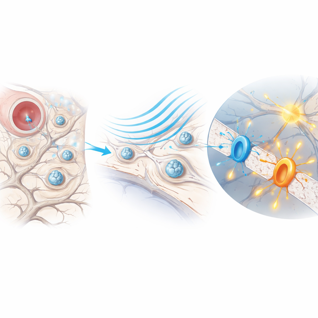
Pinpointing and Sustaining Brain Stimulation in Mice
The next step was to test the method in living mouse brains. By injecting the nanostructures into chosen regions and then applying ultrasound through the skull, the researchers could trigger muscle twitches when stimulating the motor cortex and increase activity markers only in nanostructure-filled zones deep in the striatum. Adjusting how much material they injected controlled how large the activated area became, without changing the ultrasound wavelength. Imaging showed that the particles stayed intact and functional in the brain for more than two months, providing strong ultrasound contrast and gradually fading as they were slowly cleared. Throughout this period, nerve activity in the ventral tegmental area could be repeatedly switched on with precise timing, and the pattern of activation matched where the particles had been deposited, not where sound alone might scatter.
Easing Movement Problems in Parkinson’s-Like Mice
To test therapeutic potential, the team turned to mouse models of Parkinson’s disease, in which movement becomes stiff and slow because dopamine-producing neurons in a midbrain region called the substantia nigra degenerate. They injected the hollow nanostructures into a connected relay area known as the subthalamic nucleus and applied repeated ultrasound sessions over nine weeks. In Parkinsonian mice given both particles and ultrasound, motor coordination on a rotating rod and overall movement in an open field improved steadily and remained better even after stimulation paused. Recordings from the striatum showed bursts of dopamine release precisely when ultrasound was turned on, but only in mice with nanostructures present. Brain tissue analysis revealed more surviving dopamine-producing neurons in treated mice than in those given ultrasound without particles, and a second, more chronic disease model showed similar behavioral benefits.
Safety, Limits, and Future Possibilities
The researchers carefully monitored mice for side effects. Over roughly three months, body weight, basic movement, memory, and cognition remained normal in animals that had received nanostructures alone or nanostructures plus ultrasound. Brain slices showed no clear increase in cell death or inflammation, and imaging suggested that immune cells slowly cleared the particles over time. While more work is needed to understand long-term safety, refine the materials, and adapt the method to larger brains, this study demonstrates a promising concept: by planting long-lived, sound-sensitive nanostructures once, and then stimulating them non-invasively from outside the skull, it may be possible to achieve precise, chronic control of deep brain circuits without wires, light guides, or genetic engineering.
Citation: Hou, X., Jing, J., Shi, Z. et al. Sono-mechanical nanostructures-enabled sustained precise ultrasound brain stimulation. Nat Commun 17, 3060 (2026). https://doi.org/10.1038/s41467-026-69710-8
Keywords: ultrasound brain stimulation, nanoparticles, Parkinson’s disease, neuromodulation, mechanosensitive ion channels