Clear Sky Science · en
Normative growth trajectories of fetal brain regions validated by satisfactory maturation of neurodevelopmental domains at 2 years of age
Why this matters for parents and society
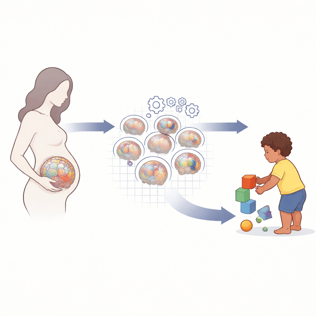
During pregnancy, the brain is the most complex organ taking shape, yet doctors usually see it only in quick snapshots on ultrasound. This study shows that with modern imaging and artificial intelligence, we can now chart how different parts of the fetal brain normally grow week by week, and link those early patterns to how children are actually developing at two years of age. These new “growth charts for the brain” could one day help identify babies at risk earlier and challenge myths about brain differences between populations.
Following thousands of pregnancies over time
The researchers drew on the INTERGROWTH‑21st Project, an international study that followed more than 4000 healthy pregnant women from Brazil, China, India, Italy, Kenya, Oman and the United Kingdom. All women were carefully selected to have good nutrition, medical care and low‑risk pregnancies, so that the study would reflect how the brain develops when conditions are close to ideal. From this group, 2805 fetuses had at least one high‑quality 3D ultrasound scan of the head between 18 and 27 weeks of pregnancy, yielding 4205 scans during this crucial mid‑pregnancy window when brain structures rapidly expand and fold.
Turning fuzzy scans into precise brain maps
Traditional methods for tracing brain structures on images require experts to outline each region by hand, which can take many hours per scan and is especially hard in ultrasound because the fetal skull casts shadows. The team instead trained deep learning algorithms to recognize and segment 16 key brain regions plus five major cortical lobes on each 3D scan. Their method uses a smart deformable model that preserves realistic brain shapes, even in shadowed areas, and can process a scan in under 10 seconds. From each scan they extracted 28 “image‑derived phenotypes” — measurements such as the total brain volume, the size of deep structures like the cerebellum and thalamus, and the thickness, depth and surface area of the developing cortex in frontal, temporal, parietal, occipital and insular lobes.
Building normal growth curves that apply across the globe
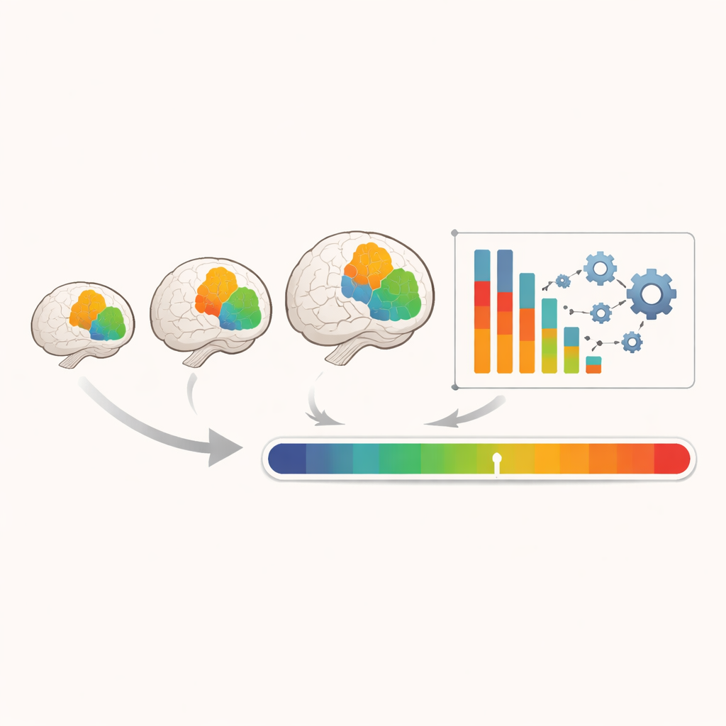
With thousands of measurements in hand, the researchers built smooth growth curves showing the 3rd, 50th and 97th percentiles for each brain region across gestational age. All regions grew quickly over the nine‑week period, but not at the same pace. When adjusted for overall brain size, many structures became relatively smaller with age, while others, such as the choroid plexus, shrank sharply in proportion. Importantly, the team checked whether brains from different study sites followed the same patterns. After accounting for sex and gestational age, differences between countries explained only 0.6% to 5.8% of the total variation for any structure, and almost all site‑by‑site comparisons fell within half a standard deviation. This means that when environmental and health conditions are good, fetal brain growth looks strikingly similar in populations with very different ancestries.
A new snapshot of brain maturity before birth
The cortex — the brain’s outer layer — does not mature uniformly. The study found that, relative to the total cortical volume, the insular lobe steadily increased in size while the parietal lobe gradually decreased between 18 and 27 weeks. The ratio of these two volumes captured this “out‑of‑step” timing of development, reflecting the closing of the Sylvian fissure and changing wiring in areas involved in body awareness, sensory integration and higher thinking. The researchers also used all 28 brain features to train a machine‑learning model that predicted gestational age from brain structure alone with an average error of about four days. This prediction serves as a “fetal brain maturation index”: if a fetus’s brain looks older or younger than its known gestational age, that difference could signal unusually fast or slow development.
Linking early brain growth to toddler abilities
To be sure that their “normal” fetal brain measurements truly corresponded to healthy outcomes, the team followed a large subset of the children to age two. Using a standardized global assessment of thinking, language, movement, behavior and vision, they excluded any children who scored in the bottom 3% for any domain. The final brain growth charts therefore reflect fetuses who later showed broadly satisfactory development. Children in this cohort, from very different world regions, achieved similar early milestones, reinforcing the finding that when social and nutritional conditions are favorable, patterns of brain growth and behavior are largely shared across humanity.
What this means for understanding early life
This work delivers the first detailed, internationally validated growth standards for multiple fetal brain regions based on fast, widely available 3D ultrasound and modern AI tools. It shows that key areas of the brain mature along predictable paths in mid‑pregnancy and that these paths look alike in well‑nourished, low‑risk populations worldwide. The new maturation index and insula‑to‑parietal ratio offer compact markers of how “on‑track” a fetal brain is, which could help future studies of high‑risk pregnancies and conditions that threaten early brain development. More broadly, the results support a powerful conclusion: differences seen between populations in brain size or child development are driven far more by unequal environments and opportunities than by inherited ancestry or skin color.
Citation: Wyburd, M.K., Kennedy, S.H., Fernandes, M. et al. Normative growth trajectories of fetal brain regions validated by satisfactory maturation of neurodevelopmental domains at 2 years of age. Nat Commun 17, 3073 (2026). https://doi.org/10.1038/s41467-026-69657-w
Keywords: fetal brain development, ultrasound imaging, deep learning, early child neurodevelopment, global health