Clear Sky Science · en
Light-induced assembly and repeatable actuation in Ca2+-driven chemomechanical protein networks
Light that makes soft matter move
Imagine a material that can grab and move tiny objects, simply because you shine light on it — and that can do this over and over again in just seconds. This paper describes such a system built from a single natural protein that shrinks when it feels a pulse of calcium. By wiring this protein to a light-sensitive calcium "switch," the authors create a soft, living-matter–inspired network that assembles, contracts, relaxes, and transports particles on command.
A springy protein borrowed from single-celled life
The work centers on Tcb2, a calcium-binding protein from the ciliate Tetrahymena, a single-celled organism covered in beating hairs. In its native context, cousins of this protein help drive some of the fastest motions in biology, where a sudden wave of calcium causes entire cells to snap into a new shape. Here, the researchers purify Tcb2 and show that, even outside the cell and without extra scaffolding, it can assemble into fibrous, web-like networks when calcium is present. Electron and fluorescence microscopy reveal that low calcium produces sparse, thin filaments, while high calcium yields dense, cortex-like sheets of protein. When calcium binds, each protein segment shortens; when calcium leaves, it lengthens again, turning the whole network into a reversible molecular spring.
Turning light into motion with a chemical relay
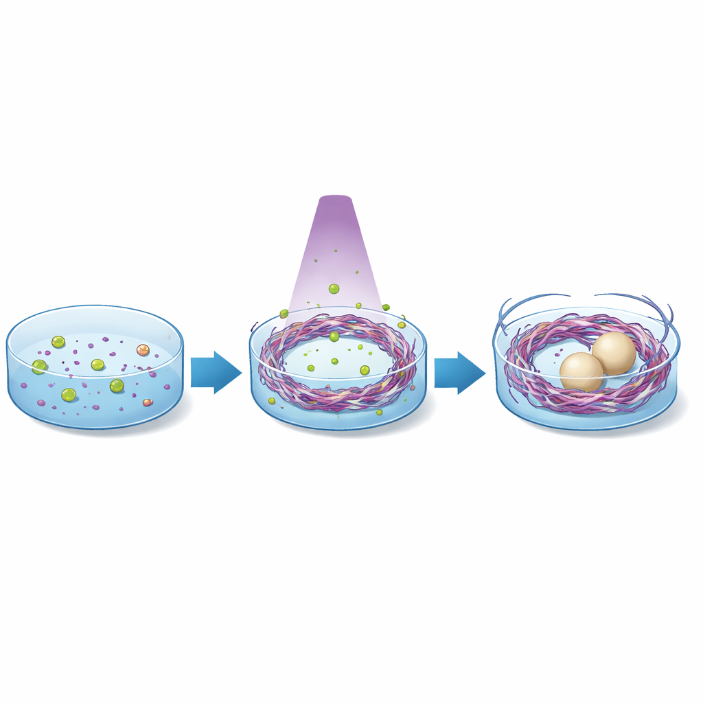
To control this spring with light, the team uses a "caged" calcium compound, DMNP-EDTA, which tightly holds calcium ions until broken by ultraviolet light. In a microscope setup with a digital micromirror device, they project patterns of 365-nanometer light into a Tcb2 solution containing the chelator and calcium. Where light hits, the chelator snaps, calcium is suddenly freed, and nearby Tcb2 proteins rapidly bind it and join the growing network. Within seconds, star-shaped or moving circular light patterns are converted into matching, contractile protein structures. A mathematical model couples how chemicals spread and react with how the soft network deforms, capturing features seen in the experiments, such as the close tracking between the spreading calcium front and the advancing edge of the protein web.
A moving ring that recharges and even reverses
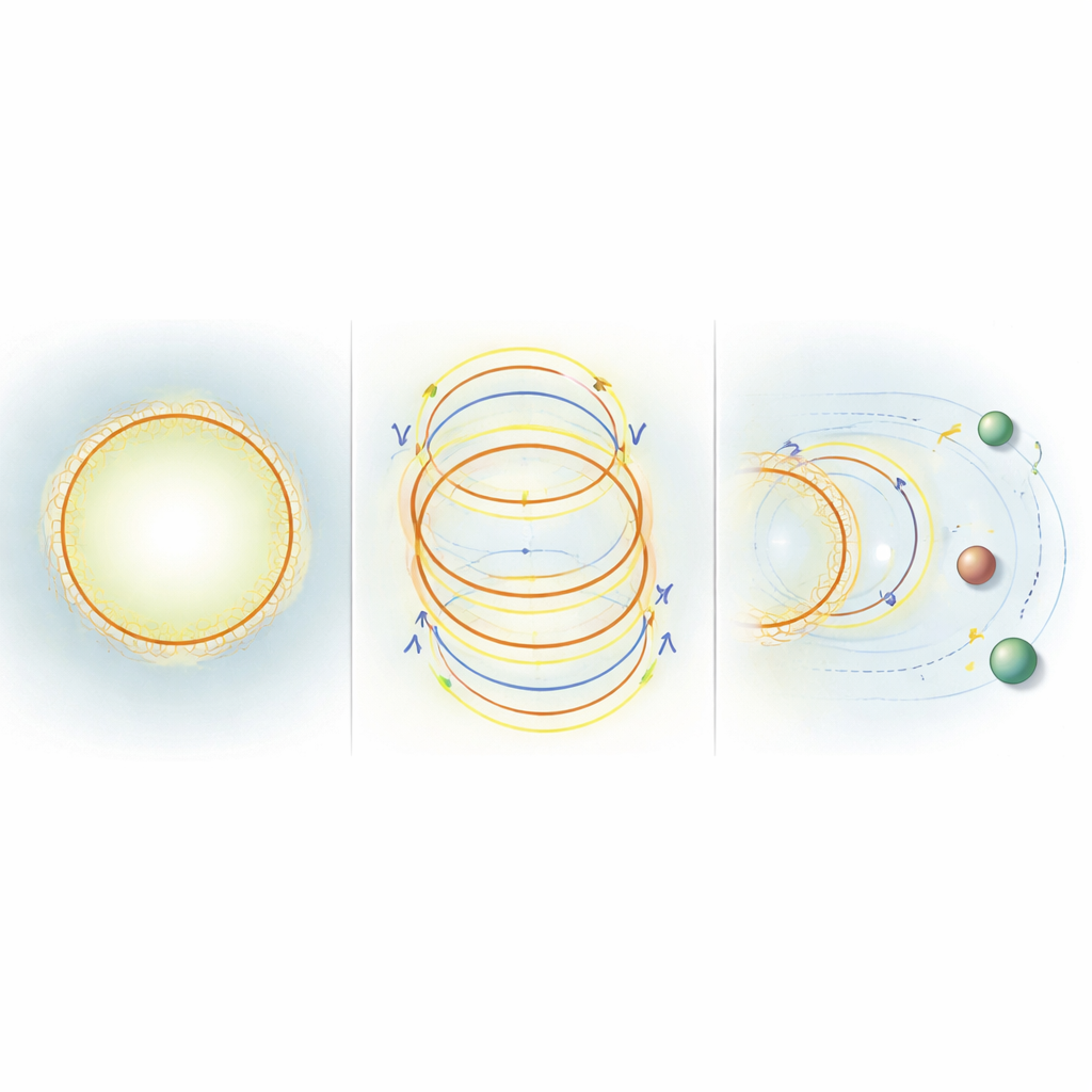
When the researchers hold a circular light spot on, the network first appears inside the lit area and then slowly grows outward as calcium diffuses away from the center. The most active zone is a narrow band at the outer edge: there, fresh Tcb2 and calcium meet, new fibers form, and the ring contracts inward like a tightening belt, while the interior remains mostly relaxed. By switching to brief light pulses separated by dark intervals, the team discovers a way to "recharge" this activity. During each pulse, calcium binds and the ring contracts; in the dark, calcium is recaptured by chelators and the fibers relax back to their longer state, but the network itself does not fully dissolve. Repeating this cycle keeps a broad region of the material mechanically lively for hundreds of rounds, and the contraction speed stays around half a micrometer per second. Surprisingly, as Tcb2 accumulates more densely near the outer boundary, the model and experiments reveal that parts of the network can briefly move outward instead of inward, because forces generated by stiffness and density gradients outweigh the simple tendency to shrink.
Using protein networks as tiny conveyor belts
Because this soft ring can push and pull, the authors test whether it can transport objects embedded in the solution. They mix in lipid vesicles and polystyrene beads and then pattern the light in space and time. Under continuous illumination, some particles caught near the forming network are drawn inward, while others farther away are shoved outward by the expanding edge, traveling tens of micrometers in seconds. With pulsed light that hops between different regions, the team can steer individual particles along more complex paths, including reversing direction multiple times as the active ring repositions. In computer simulations, they go one step further: using reinforcement learning, an algorithm learns how to adjust the radius and brightness of the light pattern so that a chosen point in the network moves to, and then holds, a target displacement. Even with only coarse feedback, the controller discovers strategies that contract quickly and then fine-tune the motion over time.
Why this matters for future soft machines
For a non-specialist, the key message is that the researchers have built a simple, programmable material that turns light into mechanical work using only three ingredients: calcium, a light-sensitive calcium holder, and a spring-like protein. Unlike many engineered systems that depend on complex motors and scaffolds, this network self-assembles fast, responds on second timescales, and can be driven repeatedly without elaborate biochemistry. The ability to draw shapes of light and have a protein web appear, pull, relax, and move attached objects hints at future uses — from deforming synthetic cell membranes to repositioning tiny organelles inside living cells. This study shows how lessons from nature’s fastest single-celled sprinters can be distilled into a controllable platform for micro-scale actuation and transport.
Citation: Lei, X., Floyd, C., Casas-Ferrer, L. et al. Light-induced assembly and repeatable actuation in Ca2+-driven chemomechanical protein networks. Nat Commun 17, 3016 (2026). https://doi.org/10.1038/s41467-026-69651-2
Keywords: light-controlled biomaterials, calcium-responsive proteins, active soft matter, microscale actuation, synthetic cytoskeletons