Clear Sky Science · en
TCF21 promotes epithelial-to-mesenchymal transition and cytoskeleton reorganization in uterine development and endometriosis
Why this research matters for women’s health
Endometriosis affects millions of women worldwide, causing chronic pain, heavy periods, and infertility, yet its roots inside the body remain surprisingly mysterious. This study uncovers how a single gene regulator, called TCF21, shapes the womb during early life and later helps drive the spread and persistence of endometriosis. By connecting normal uterine development with a common disease, the work points to new ways to diagnose, prevent, and possibly treat endometriosis by targeting the same molecular pathway.
Building the inner lining of the womb
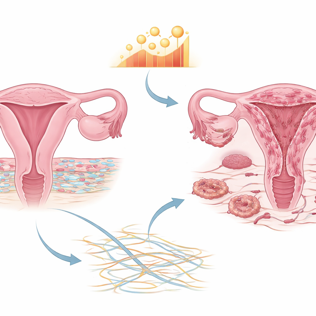
The womb’s inner lining, the endometrium, is not a uniform sheet of cells. It is a carefully layered tissue in which a surface “skin” of epithelial cells sits atop a supportive bed of stromal cells. In newborn mice, this structure is still forming. The researchers showed that TCF21 is switched on just as the uterus is maturing after birth, peaking around the time when glands and the supporting stroma are being built. When they removed the Tcf21 gene specifically from the uterus in mice, the animals developed unusually thin endometrial linings with far fewer stromal cells, even though their hormones, ovaries, and ability to ovulate and fertilize eggs were largely normal. These mice were much less fertile, producing fewer litters and fewer pups per litter.
When cells change identity and go rogue
To understand why stromal cells were missing, the team focused on a process called epithelial-to-mesenchymal transition, in which orderly, stationary epithelial cells gradually acquire traits of more mobile, flexible stromal cells. In healthy uterine development, this transition helps generate the stromal compartment. In the Tcf21-deficient mice, key markers of stromal identity were reduced while epithelial markers increased, pointing to a stalled transition. In women with endometriosis, by contrast, the picture was reversed: samples from normal womb lining, the lining inside the uterus of women with endometriosis, and the abnormal tissue growing outside the uterus showed a stepwise rise in stromal cells and in TCF21 levels. Single-cell analyses confirmed that in ectopic lesions—patches of endometriosis in places like the ovary—stromal-like cells dominate and TCF21 is especially abundant in their nuclei.
How a gene regulator reshapes the cell’s internal scaffolding
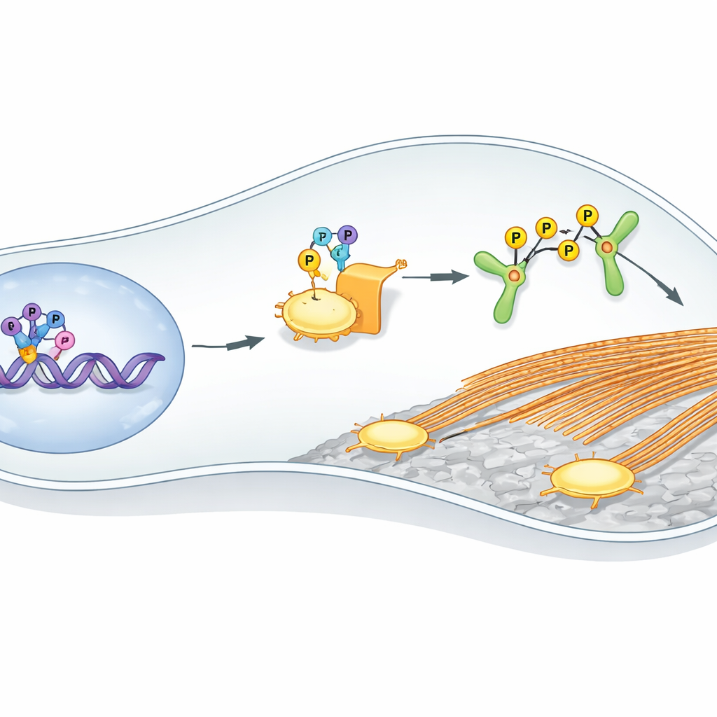
Having linked TCF21 to changes in cell identity, the researchers asked how it might make stromal cells more invasive. They mapped TCF21’s docking sites across the genome in human endometriotic stromal cells and combined this with gene activity data from patient tissues. A striking pattern emerged: many TCF21 targets control the cell’s internal scaffolding—the actin cytoskeleton—and the structures that allow cells to grip and pull on their surroundings, called focal adhesions. One key target was LIMK2, an enzyme that modifies a protein named cofilin, which normally helps break down actin filaments. When TCF21 levels were raised in stromal cells, LIMK2 and activated cofilin increased, actin filaments became more abundant and organized, and focal adhesions grew in number and size. Lowering TCF21 had the opposite effect, and manipulating LIMK2 could reverse these changes, showing that TCF21 drives a LIMK2–cofilin pathway that stiffens the cell’s skeleton and boosts its ability to move and stick.
From petri dish to living animals
The team then tested whether this pathway actually fuels disease in living animals. In a mouse model where bits of uterine lining are surgically grafted into the abdomen to mimic endometriosis, animals lacking uterine Tcf21 developed smaller and fewer lesions than normal mice. These lesions also showed weaker LIMK2–cofilin signaling. In a complementary experiment, the researchers used a tailored virus to boost Tcf21 specifically in the uterus of mice. Those animals developed larger endometriosis-like lesions, but when they were treated with a small-molecule drug that blocks LIM kinases, lesion growth was sharply reduced—even in the presence of high Tcf21. Importantly, patient samples echoed these animal results: in paired tissues from women with endometriosis, both TCF21 and LIMK2 were consistently higher in ectopic lesions than in the lining inside the uterus and their levels rose and fell together.
What this means for future diagnosis and treatment
This study paints a coherent picture: TCF21 is a master switch that helps build a healthy uterine lining in early life, but when its activity is abnormally high later on, it nudges cells toward a more mobile, invasive state. By cranking up LIMK2 and reorganizing the actin scaffold inside stromal cells, TCF21 makes it easier for fragments of endometrial tissue to latch onto new sites in the pelvis and dig in, contributing to endometriosis. Because directly blocking a transcription factor like TCF21 is technically challenging, the LIMK2–cofilin arm of the pathway offers a more practical target. Drugs that safely dampen this signaling could, in principle, slow lesion growth and reduce pain and infertility in women with endometriosis, while measurements of TCF21–LIMK2 activity might help identify those at higher risk or track how well treatments are working.
Citation: Zhu, J., Wu, P., Ma, Y. et al. TCF21 promotes epithelial-to-mesenchymal transition and cytoskeleton reorganization in uterine development and endometriosis. Nat Commun 17, 3420 (2026). https://doi.org/10.1038/s41467-026-69551-5
Keywords: endometriosis, uterine development, TCF21, cell cytoskeleton, LIMK2