Clear Sky Science · en
Multi-lineage hepatic organoids reveal toxic exosome mediated indirect hepatotoxicity
Why this matters for medicines and liver health
Many promising medicines never reach patients, or are later withdrawn, because they unexpectedly damage the liver. Some of the trickiest cases are drugs that do not poison liver cells directly but instead set off a slow chain reaction involving supporting cells and microscopic messengers. This study introduces a lab-grown mini-liver model that mimics these complex cell-to-cell conversations and uses it to uncover how a common antidepressant can quietly injure the liver through a hidden, indirect route.
Building a mini-liver in a dish
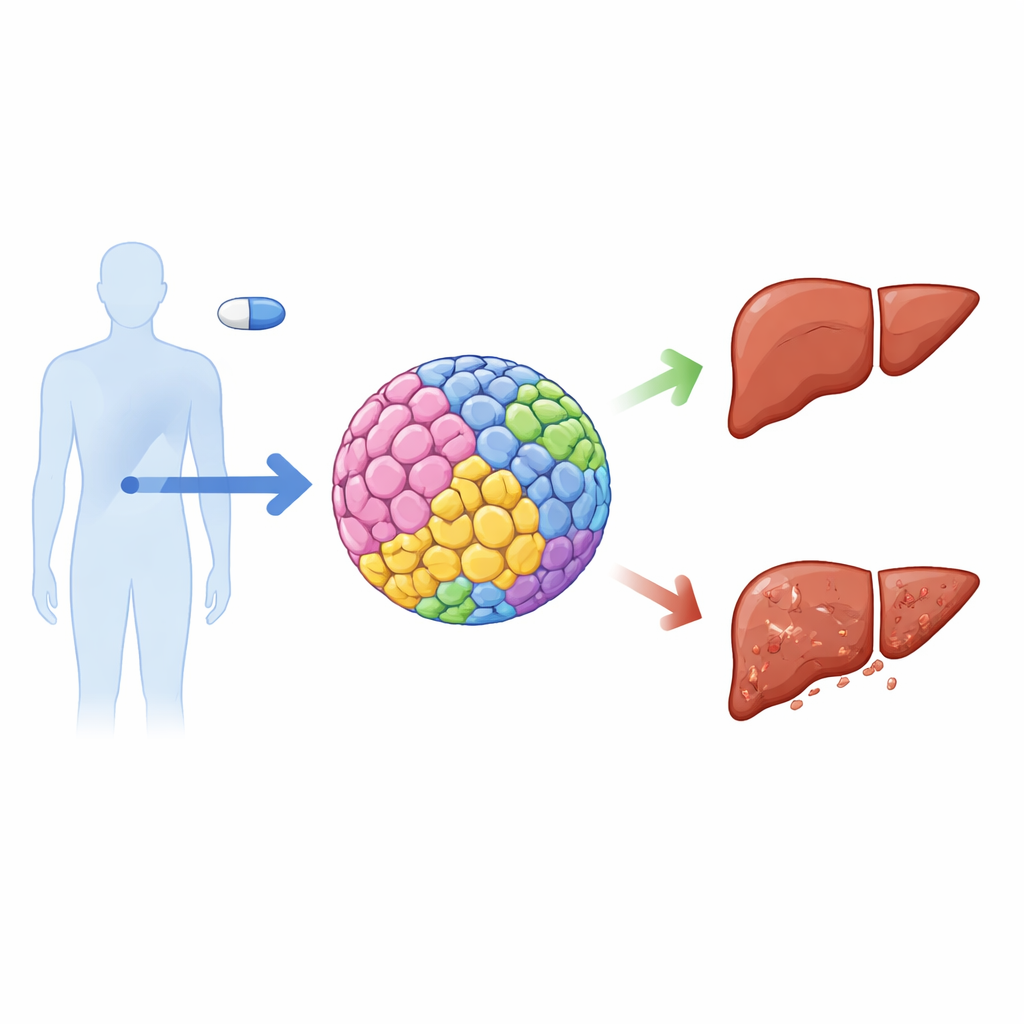
The liver is not made of a single cell type, yet most safety tests rely on flat layers of only one kind of liver cell. The authors created three-dimensional "hepatic organoids"—tiny spheres that mimic the architecture of real liver tissue. Starting from human embryonic stem cells, they guided development into five key liver cell types: drug-processing hepatocytes, bile duct cells, blood vessel lining cells, immune-like Kupffer cells, and scar-forming stellate cells. These cells were mixed in realistic ratios and grown into stable spheroids that maintained structure and function for over a month, secreting albumin, processing nutrients, and responding strongly to inflammatory signals.
When the researchers compared these multi-cell organoids to simpler models, they found that the organoids most closely matched human liver tissue at the gene-expression level and showed the highest activity of drug-metabolizing enzymes. They also handled tasks such as urea production, fat handling, and sugar storage better than standard two-dimensional cultures or hepatocyte-only spheroids. This suggested that the added cell types and three-dimensional layout recreated important aspects of the liver’s internal environment, making the organoids a more realistic testing ground for drug safety.
Finding drugs that harm the liver indirectly
To test how useful this model is for predicting real-world liver injury, the team exposed the organoids to 58 drugs with known clinical records—some safe for the liver, others known to cause damage. Each drug was tested at a concentration similar to levels seen in patients, over two weeks. The organoids correctly flagged most of the harmful drugs and correctly spared most of the safe ones, achieving over 80% sensitivity and 75% specificity. Intriguingly, several drugs appeared toxic only in the multi-cell organoids, not in hepatocyte-only spheroids, hinting at injury driven by cross-talk among cell types rather than direct poisoning of hepatocytes.
One antidepressant, imipramine, stood out. In the complex organoids it caused clear loss of viability, yet it did not directly kill isolated hepatocytes. Follow-up experiments pointed to stellate cells as the key responders: they carry high levels of a receptor called TRKB that can bind imipramine. When stellate cells with normal TRKB were exposed to the drug, they underwent marked stress and cell death, whereas stellate cells with TRKB knocked down were largely protected. This suggested that the drug acts first on stellate cells, which then send damaging signals onward to neighboring hepatocytes.
Toxic messages in tiny vesicles
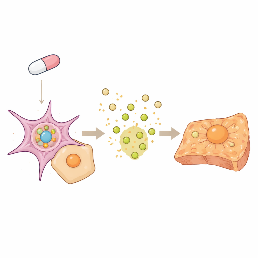
The researchers suspected that exosomes—nanoscale bubbles that cells use to ship molecular cargo—might carry the harmful signal. They collected exosomes from stellate cells treated with imipramine and showed that these vesicles were readily swallowed by hepatocytes. Blocking exosome uptake with an antibody reduced this transfer. Importantly, the number and size of vesicles did not change with drug treatment; instead, their contents did. Hepatocytes that received exosomes from drug-treated stellate cells switched on cell-death machinery and turned down XIAP, a protein that normally prevents apoptosis.
Further molecular detective work traced this change back to a small regulatory RNA, miR-34a-3p. Imipramine, acting through TRKB in stellate cells, activated a pathway involving p53 and RNA-processing proteins that selectively enriched miR-34 in the exosomes. This microRNA directly targets XIAP in hepatocytes, tipping the balance toward activation of caspase-3 and programmed cell death. When mice were given imipramine over several weeks, the same pattern appeared: stellate cells showed early activation of p53 and miR-34, exosome markers accumulated around hepatocytes, and hepatocyte death and blood liver enzymes rose later on. Interfering with TRKB, exosome formation, or miR-34 in vivo all reduced liver injury.
What this means for future drug safety
This work shows that a carefully engineered mini-liver can reveal subtle, indirect forms of drug-induced liver injury that conventional tests may miss. In the case of imipramine, the drug does not primarily harm hepatocytes on its own. Instead, it disturbs stellate cells, which in turn send out toxic exosomes loaded with miR-34 that silence a key survival protein in hepatocytes and trigger their death. By capturing this multi-step communication, the organoid model not only improves early safety screening but also points to new ways to prevent damage—such as blocking harmful exosome cargo—before problems appear in patients.
Citation: Sun, L., Zhang, Y., Niu, Y. et al. Multi-lineage hepatic organoids reveal toxic exosome mediated indirect hepatotoxicity. Nat Commun 17, 2926 (2026). https://doi.org/10.1038/s41467-026-69548-0
Keywords: drug-induced liver injury, liver organoids, exosomes, imipramine, microRNA-34