Clear Sky Science · en
Single-cell multiomic human brain atlas reveals regulatory drivers of cortical regionality
Why brain maps are getting a powerful upgrade
Different parts of the human brain’s outer layer, the cortex, handle everything from seeing and hearing to planning, language, and social thought. But what actually makes one patch of cortex behave differently from another? This study builds a detailed atlas of the human cortex at single-cell resolution, measuring not only which genes are active in millions of individual cells, but also how the DNA around those genes is opened or closed. By tying these molecular patterns to precise locations in the brain, the work reveals hidden control circuits that tune brain regions for their specialized roles and may shape vulnerability to disorders such as autism and Alzheimer’s disease.
Looking closely at many tiny brain parts
The researchers analyzed tissue from nine cortical regions that span a broad range of functions, including areas for movement, sensation, hearing, vision, and higher-order thinking. From postmortem brains of six donors, they isolated more than three million cell nuclei and used a dual-omics method to read, in the same cells, both RNA (which genes are turned on) and chromatin accessibility (which stretches of DNA are open for regulatory proteins to bind). They also used a spatial imaging method to map the positions of about 157,000 cells directly in tissue slices. Combining these approaches produced a rich “multiomic” atlas that connects cell identity, molecular state, and physical location across the cortex. 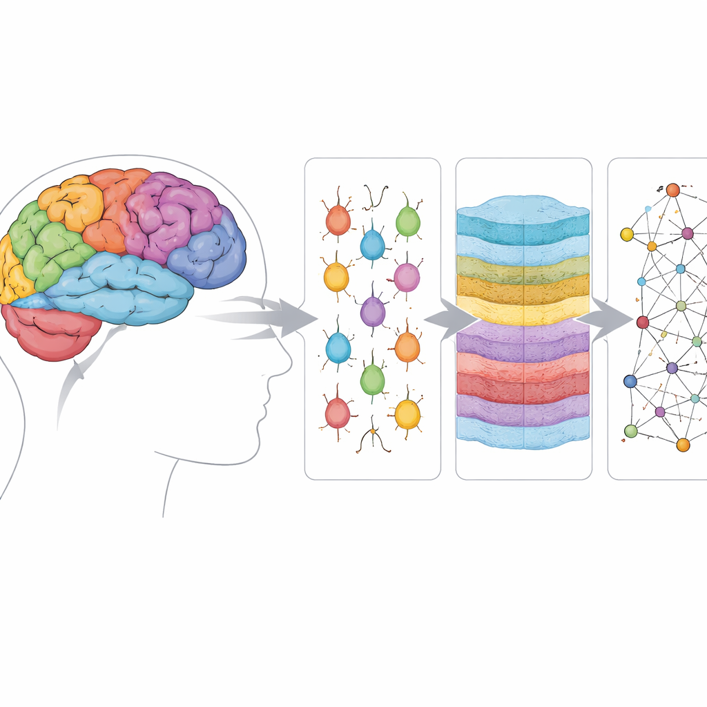
Finding the brain’s main cell players
By clustering the molecular profiles, the team identified 24 broad subclasses and 120 finer cell types, including several kinds of excitatory and inhibitory neurons as well as non-neuronal support cells. The clearest regional differences appeared in intratelencephalic (IT) projection neurons—cells that send signals to other cortical areas—and in certain deep-layer and inhibitory neurons. The authors cataloged thousands of genes whose activity varies by region and showed that many of these genes relate to how neurons grow, wire up, and communicate. They also mapped hundreds of thousands of candidate regulatory DNA elements and linked them to their likely target genes, revealing region- and cell-type–specific control switches embedded in the genome.
Hidden gradients that run across the cortex
Instead of treating each region as an island, the team asked how molecular patterns change smoothly along known axes that organize the cortex. Along a front-to-back (rostral–caudal) direction, IT neurons, especially those in layer 4, showed striking shifts in genes that manage calcium levels inside cells and translate electrical activity into long-term changes. Key components of calcium entry, pumping, and downstream signaling all varied systematically across this axis. A second axis separated sensory regions (visual, auditory, somatosensory, motor) from “transmodal” association areas that integrate information. Along this transmodal–sensory axis, the researchers saw coordinated “subunit switching” in major receptor families: different molecular building blocks of the same receptor were favored in different regions, subtly altering how neurons respond to glutamate, GABA, acetylcholine, and serotonin.
Control circuits behind regional specialization
To move beyond lists of genes, the authors inferred gene regulatory networks—who controls whom—by combining chromatin and expression data. They pinpointed transcription factors whose binding activity and own expression change in step with cortical axes, and whose predicted target genes follow the same gradients. For calcium-related genes in layer-4 IT neurons, a small group of such factors, including BACH2, KLF12, and TCF12, emerged as key regulators. For receptor subunit switching along the transmodal–sensory axis, factors like RFX3 and TCF4 stood out, with regulatory DNA near important receptor genes such as GRIN2B showing strong predicted binding. Notably, many of these regulators have been implicated in autism and other neurodevelopmental conditions, suggesting that disruptions in these finely tuned gradients could help explain why certain regions are especially affected. 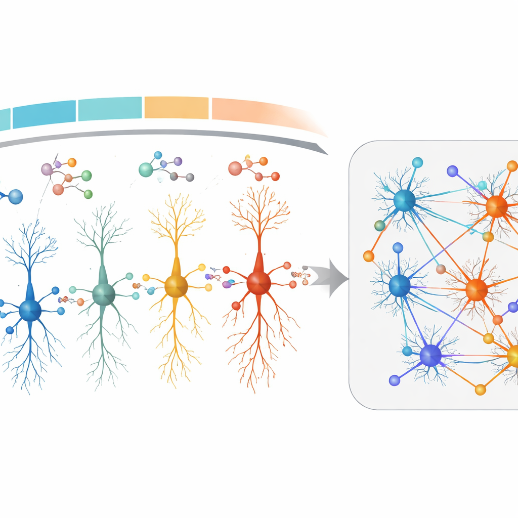
What this means for understanding brain health
In plain terms, this work shows that the cortex is not just divided into different areas by anatomy or connectivity; it is also sculpted by smoothly varying molecular programs that tweak how similar types of neurons behave from place to place. These programs adjust how easily neurons fire, how they respond to key chemical signals, and how they store information over time, thereby helping each region meet its particular demands. Because the same regulatory networks that shape normal regional specialization also intersect with genes tied to autism and Alzheimer’s disease, this atlas offers a roadmap for exploring why some circuits are fragile and others are resilient. It provides a foundational reference for linking microscopic gene control to large-scale brain function and dysfunction.
Citation: Palmer, C.R., Song, J., Yang, B. et al. Single-cell multiomic human brain atlas reveals regulatory drivers of cortical regionality. Nat Commun 17, 3051 (2026). https://doi.org/10.1038/s41467-026-69368-2
Keywords: human cortex, single-cell multiomics, gene regulatory networks, brain regionalization, neurodevelopmental disorders