Clear Sky Science · en
Conformational biosensors delineate endosomal G protein regulation by GPCRs
How Cells Send Second-Wave Signals Inside
Many medicines work by targeting receptors on the surface of our cells, but scientists now know that these receptors can keep sending messages from deep inside the cell as well. This study asks a deceptively simple question: once a surface receptor is switched on, how does the cell make sure the right internal compartments receive a fresh supply of signaling molecules, and how does it decide which kind of signal to send from those inner stations?
From Surface Switches to Inside Compartments
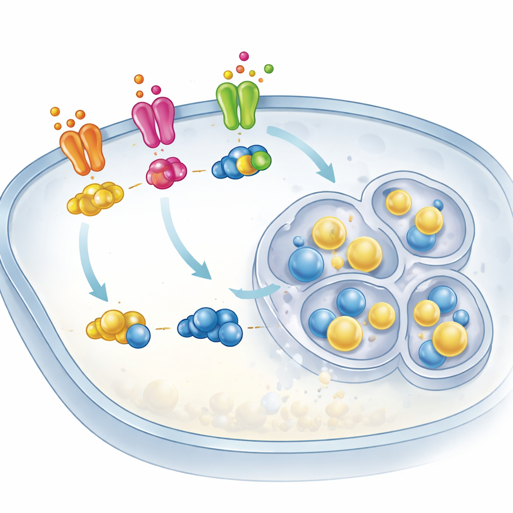
The receptors in this story belong to the huge family of G protein–coupled receptors, or GPCRs, which respond to hormones, neurotransmitters, and many drugs. When activated at the cell’s outer membrane, these receptors turn on helper proteins called G proteins that carry the signal inward. The authors focus on one widespread G protein type, often called Gs, which normally sits at the cell surface but has been suspected to move to internal bubbles of membrane known as endosomes, where it can launch a second wave of signaling. Until now, however, scientists had not directly seen Gs in its active, signal-carrying form on these endosomes or understood how it got there.
Tracking Moving Parts in Living Cells
To watch this process unfold in real time, the team built sensitive molecular “biosensors” that light up only when G proteins adopt specific shapes. One biosensor detects Gs after it has bound the energy-rich molecule that marks it as active, while another recognizes G proteins as they are being engaged by a receptor. Using advanced microscopy and light-emitting readouts in human cells, the researchers followed where Gs traveled and when it turned on. They examined three different receptors that all stimulate Gs but have distinct behaviors: the classic beta-2 adrenergic receptor, a receptor for the gut hormone vasoactive intestinal peptide, and an adenosine receptor involved in immune and vascular control.
Two Separate Steps: Getting There and Turning On
The experiments revealed that activation of any of these receptors at the cell surface quickly causes Gs to detach from the outer membrane and spread through the cell, including onto endosomes. Surprisingly, this reshuffling did not require the receptors themselves to be dragged inside; blocking receptor entry into the cell did little to stop Gs from relocating. However, the story changed when the team asked where Gs actually became active. Here, the biosensors showed a clear two-phase pattern: first, Gs was activated at the plasma membrane; then, after a delay, active Gs accumulated on endosomes. Crucially, this second phase depended on receptors being physically present on endosomes. When endocytosis—the process that pulls receptors inward—was inhibited, the rise of active Gs on endosomes dropped sharply even though Gs still arrived there.
Where You Signal Changes What You Say
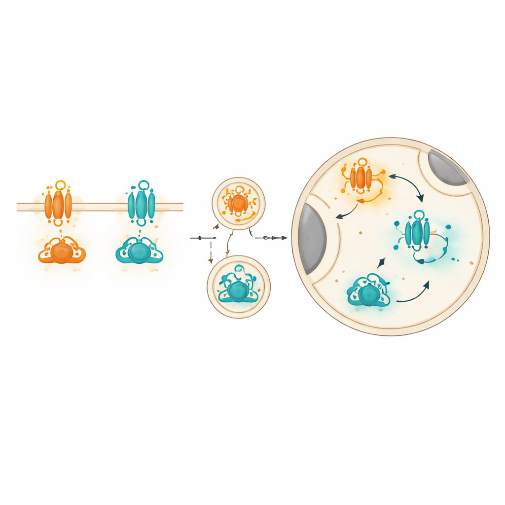
GPCRs can talk not only to Gs but also to other G protein types, such as Gq, which trigger different cellular responses. By carefully dissecting biosensor signals, the authors found that one receptor (VIPR1) activates both Gs and Gq at the cell surface but mainly Gs on endosomes. In contrast, another receptor (A2B) activates both Gs and Gq at the surface yet favors Gq on endosomes, even though it barely enters endosomes itself. This “location bias” means that the same receptor can send a different mix of signals depending on whether it is at the edge of the cell or inside it, and that different receptors imprint their own characteristic balance of G proteins in each location.
Why This Matters for Medicines
For a non-specialist, the key message is that cell-surface receptors do not just turn signals on or off; they also control where in the cell those signals originate and which flavor they take. This work shows that one step at the surface moves G proteins inward, while a second, separate step on endosomes turns them on again. It also shows that different receptors bias this internal signaling toward different G protein types. These insights help explain why drugs acting on the same receptor family can have subtly different effects—and suggest that future medicines might be designed to steer signaling not only toward particular pathways, but also toward specific locations inside the cell.
Citation: Wysolmerski, B., Fisher, N.M., Dates, A.N. et al. Conformational biosensors delineate endosomal G protein regulation by GPCRs. Nat Commun 17, 2911 (2026). https://doi.org/10.1038/s41467-026-69329-9
Keywords: GPCR signaling, endosomal signaling, G protein trafficking, cellular biosensors, location-biased drugs