Clear Sky Science · en
Sex-specific differences in mediobasal hypothalamus in response to nutritional states
Why this research matters for everyday health
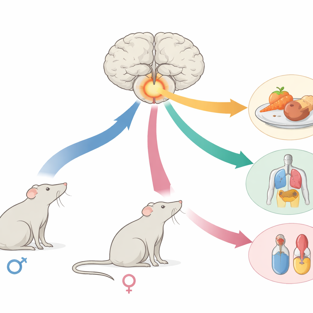
Obesity, diabetes, and related metabolic diseases affect hundreds of millions of people, yet men and women often gain weight differently, store fat in different places, and respond differently to diets. This study asks a basic but overlooked question: inside the brain’s appetite and hormone centers, do male and female cells actually respond differently to being well fed or very hungry? Using a powerful gene-reading technique in mice, the researchers map how thousands of individual brain cells react to feeding and fasting in each sex, offering clues to why metabolism and fertility are so strongly shaped by both nutrition and biological sex.
A small brain hub with wide-reaching influence
The work focuses on a tiny region deep in the brain called the arcuate nucleus, part of the mediobasal hypothalamus. Despite its small size, this hub helps decide when we feel hungry, how we burn or store energy, how we grow, and when we are ready to reproduce. The same region also shows clear differences between males and females in many species. To probe its inner workings, the authors examined more than 90,000 individual cell nuclei from male and female mice that were either allowed to eat freely or fasted for 28 hours, a period chosen to ensure the animals were strongly motivated to seek food.
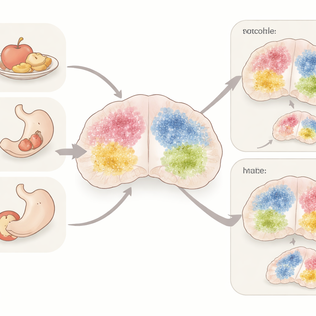
Taking a cell-by-cell look at hunger and sex
By reading RNA—the working copies of genes—from single nuclei, the team grouped cells into 42 distinct types, including 31 types of neurons and 11 types of support cells. They then asked, for each cell type, which genes turned up or down with fasting, and which differed between males and females. The most striking changes appeared in a set of hunger-driving neurons called Agrp cells, which were strongly activated by fasting in both sexes. Another population, Pomc neurons, which tend to curb eating, also showed meaningful but more modest shifts with nutritional state. Importantly, the researchers confirmed that these patterns were not technical artifacts by carefully correcting for batch effects and comparing their data to earlier brain atlases.
How male and female brains diverge
Some neuron groups were especially sensitive to sex, nutrition, or both. KNDy neurons, which help control reproductive hormone pulses, showed dramatic differences between females and males and responded strongly to fasting only in females. Dopamine-producing neurons in the same region were also highly sex-specific and changed with nutrition mainly in females. Many of the genes that differed between sexes were located on non–sex chromosomes, indicating that sex hormones and lifelong hormonal history, rather than just XX or XY status, likely shape these patterns. In contrast, most support cells, such as microglia and oligodendrocytes, remained relatively stable, though they showed subtle gene changes hinting at shifts in inflammation and nerve insulation during fasting.
Signals that link hunger, wiring, and hormones
Because many of the changing genes were related to cell-to-cell signaling, the scientists modeled how different cell types might be “talking” to one another. They found that neurotrophic factors—molecules that support growth and connectivity of neurons—were key messengers tuned by both sex and nutrition. During fasting, hunger-promoting Agrp neurons in females ramped up certain neurotrophic signals, while satiety-related Pomc neurons dialed them down. Reproductive and dopamine neurons in females also showed higher levels of related signals and receptors than in males. These patterns suggest that prolonged hunger not only changes immediate activity in appetite circuits but may also reshape their wiring over time, in ways that differ between male and female brains.
What this means for future treatments
Overall, the study shows that the brain’s core appetite and hormone center does not respond to hunger and plenty in a uniform way. Instead, specific neuron types tune their gene activity differently depending on sex and nutritional state, with female cells often showing stronger and more complex changes. Support cells participate too, but more quietly. For a layperson, the key message is that male and female brains manage energy balance and reproduction with overlapping but distinct cellular programs. This deeper map of sex- and diet-sensitive cells in the hypothalamus could guide the design of future obesity and metabolic therapies that are better matched to biological sex, improving both effectiveness and safety.
Citation: Bean, J.C., Jian, J., Lu, TC. et al. Sex-specific differences in mediobasal hypothalamus in response to nutritional states. Nat Commun 17, 2941 (2026). https://doi.org/10.1038/s41467-026-69239-w
Keywords: hypothalamus, sex differences, fasting, energy balance, single-cell RNA sequencing