Clear Sky Science · en
RNA-binding proteins Zfp36l1 and Zfp36l2 protect against premature thymic involution
Why This Matters for Your Immune System
The thymus is a small organ tucked above the heart that quietly shapes our immune defenses, especially in childhood. As we age, this organ naturally shrinks, making it harder to produce new T cells that fight infections and respond to vaccines. This study explores why the thymus wears out over time and identifies two tiny RNA-binding proteins, Zfp36l1 and Zfp36l2, as key guardians that help keep the thymus healthy by preventing harmful inflammation inside it. 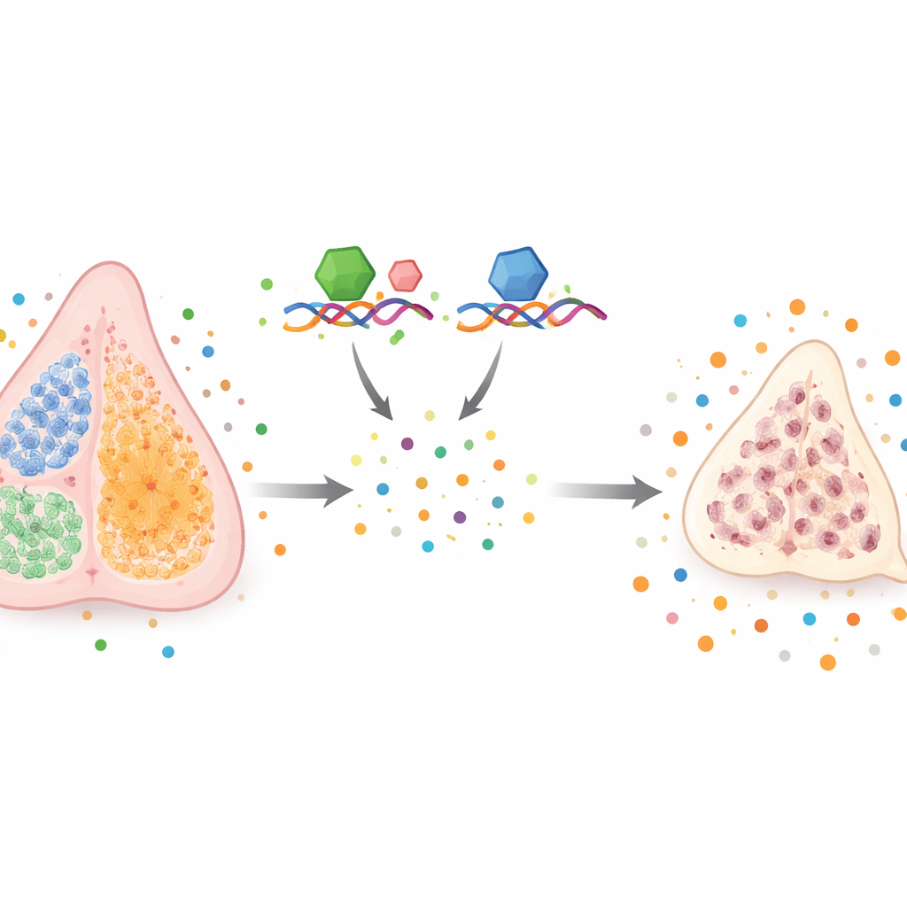
The Thymus: A Training School Under Stress
The thymus acts like a specialized school where immature immune cells learn to recognize germs while ignoring the body’s own tissues. This education depends on thymic epithelial cells, or TECs, which come in two main varieties: cortical TECs (teachers for early lessons) and medullary TECs (teachers for self-tolerance, preventing autoimmunity). With age or under chronic stress, the thymus shrinks and its output of fresh T cells drops. Earlier work showed that a gene called Foxn1 is crucial for keeping TECs healthy and that chronic inflammatory molecules, such as interferons and interleukin-6, can accelerate thymic shrinkage. However, what controls the levels of these inflammatory signals inside the thymus itself has been unclear.
RNA Guardians That Keep Signals in Check
The proteins Zfp36l1 and Zfp36l2 belong to a family that binds to specific RNA sequences and flags those RNAs for destruction, acting as post-transcriptional brakes. Many of their targets are messages that encode inflammatory cytokines or cell-cycle regulators. The researchers engineered mice in which Zfp36l1 and Zfp36l2 were removed only from TECs, leaving the rest of the immune system intact. They expected this might actually slow thymic aging by stabilizing certain helpful RNAs, but instead observed the opposite: by three weeks of age, and even more dramatically by adulthood, these mice had much smaller, cell-poor thymuses, resembling prematurely aged organs.
Early Cell Loss and Skewed Thymic Architecture
Closer examination showed that TEC numbers started falling before birth, even when the total size of the thymus still looked normal. Early on, cortical TECs were preferentially lost, while medullary TECs became relatively more abundant. Single-cell RNA sequencing revealed that TECs lacking Zfp36l1 and Zfp36l2 were skewed toward immature medullary-like states and showed increased expression of genes that slow the cell cycle, such as p21 and p57. This suggests that without these RNA-binding proteins, TECs divide less efficiently and their normal maturation pathways are disrupted. 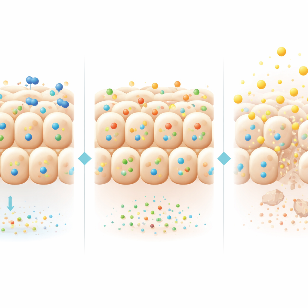
A Tug-of-War Between Growth and Inflammation
The study uncovered a striking time-dependent shift in Foxn1, the master gene that maintains TEC health. During embryonic life, Foxn1 protein levels were actually higher in the remaining mutant TECs, likely because its RNA was no longer being degraded efficiently. This temporary boost may help sustain thymus function despite fewer TECs. But after birth, as the thymus enters a period of rapid growth and medullary TECs begin to produce more inflammatory cytokines, Foxn1 levels in the mutant TECs fell below normal. At the same time, medullary TECs without Zfp36l1 and Zfp36l2 produced abnormally high amounts of inflammatory cytokines such as interleukin-6 and tumor necrosis factor. Organ culture experiments showed that thymic stromal tissue from these mutants collapsed over time, and even when mixed with normal tissue, the mutant environment drove deterioration, underscoring a harmful, self-sustaining inflammatory loop.
Impacts on Immune Cell Education
This altered thymic environment affected not only TECs but also the immune cells they educate. The composition and activation of dendritic cells and B cells inside the thymus shifted, and the maturation of certain T cell subsets, particularly CD8 T cells, was impaired during early life. Although blocking a few individual cytokines, including type I interferons, interleukin-6, and tumor necrosis factor, was not enough to rescue the thymus, the data indicate that a broader mix of misregulated inflammatory signals contributes to both TEC stress and flawed T cell development.
What This Means for Thymus Aging
Overall, the study shows that Zfp36l1 and Zfp36l2 act as internal safety valves in thymic epithelial cells, fine-tuning the levels of many inflammatory messages and helping to maintain Foxn1 and TEC health. When these RNA-binding proteins are lost, TECs become fewer and more dysfunctional, inflammatory cytokines rise, Foxn1 eventually declines, and the thymus shrinks far earlier than it should. For a layperson, this suggests that healthy aging of the immune system depends not only on whether the right genes are turned on, but also on how quickly their RNA messages are cleared away. Understanding and potentially mimicking the action of these RNA guardians could one day help preserve thymus function, sustain T cell production, and improve immune resilience in older adults.
Citation: Han, J., Golzari-Sorkheh, M., Rajan, V. et al. RNA-binding proteins Zfp36l1 and Zfp36l2 protect against premature thymic involution. Cell Mol Immunol 23, 505–516 (2026). https://doi.org/10.1038/s41423-026-01399-7
Keywords: thymus aging, RNA-binding proteins, thymic epithelial cells, inflammatory cytokines, T cell development