Clear Sky Science · en
B cells maintain the homeostasis of splenic marginal zone antigen-presenting cells to promote the antiviral CD8+ T-cell response
Why our own defenders need quiet helpers
Most of us have heard that killer T cells are frontline warriors against viruses, especially in people whose immune systems are weakened by transplants or disease. But this study shows that another immune cell type, B cells—best known for making antibodies—quietly sets the stage for those killer T cells to work. By uncovering how B cells help organize a critical region of the spleen, the authors reveal a hidden support network that could influence how we treat viral infections, immune disorders, and even some cancers.
A busy border station inside the spleen
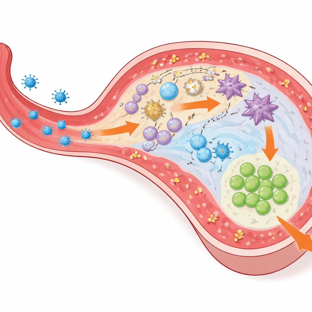
The spleen acts as a major filter for our blood, catching germs as they circulate. At the edge of its white, immune-rich tissue lies a narrow ring called the marginal zone. This zone is packed with different sentry cells: special macrophages that grab passing virus, dendritic cells that show viral pieces to T cells, and B cells that usually make antibodies. When a blood-borne virus such as cytomegalovirus (CMV) arrives, its first real encounter with the immune system often happens here. The authors used a mouse version of CMV to dissect how these cells cooperate to launch a strong CD8+ T-cell attack, which is crucial for keeping CMV in check, especially in vulnerable patients.
When B cells are missing, killer T cells fall behind
The team first examined mice that completely lacked B cells. After CMV infection, these mice produced far fewer virus-specific CD8+ T cells and allowed more viral activity, even though their remaining CD8+ T cells were fully capable of attacking infected cells. To test whether antibodies were to blame, the researchers used mice that had B cells but could not secrete normal antibodies, and also transferred serum containing CMV antibodies into B-cell-deficient animals. In both cases, the CD8+ T-cell response was restored or remained normal despite defective antibodies, showing that the key role of B cells here was not to make antiviral proteins, but something more structural and local within the spleen.
Specialized dendritic cells need a stable neighborhood
Looking deeper, the scientists focused on dendritic cells, the immune “interpreters” that prime CD8+ T cells. They found that a particular dendritic subset, called Langerin+ cDC1, was greatly reduced in mice without B cells. These dendritic cells sit in the marginal zone and are especially good at taking up viral material and driving robust CD8+ T-cell activation. When the researchers experimentally removed Langerin+ dendritic cells in otherwise normal mice, the CMV-specific CD8+ T-cell response dropped, mirroring what they saw in B-cell-deficient animals. This pointed to a simple chain: fewer Langerin+ dendritic cells meant weaker priming of antiviral T cells.
How B cells keep the marginal zone in shape
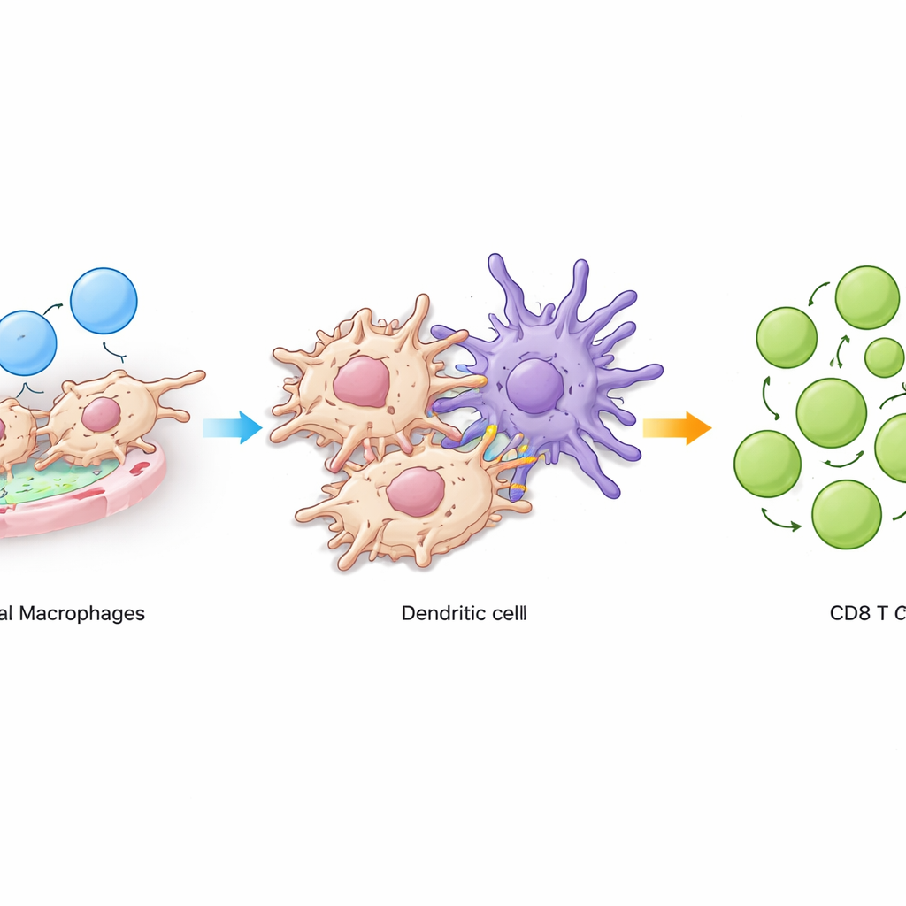
The next question was how B cells support these dendritic cells. The authors showed that B cells produce a signal called lymphotoxin β, which is needed to maintain a particular group of macrophages in the marginal zone known as CD169+ metallophilic macrophages. These macrophages cluster at the border where blood first enters the spleen and physically interact with Langerin+ dendritic cells. Using tracking experiments, the team demonstrated that macrophages pass captured material to these dendritic cells, allowing them to present viral pieces efficiently to CD8+ T cells. When B cells lacked lymphotoxin β, the marginal-zone macrophages declined, Langerin+ dendritic cells shrank in number, and the antiviral CD8+ T-cell response weakened.
A molecular handshake that holds the circuit together
Single-cell genetic analysis and imaging then highlighted a key molecular “handshake” between macrophages and Langerin+ dendritic cells. Macrophages expressed an adhesion molecule called VCAM1, while the dendritic cells expressed a matching integrin receptor. These molecules formed tight contact points where the two cell types touched. Blocking VCAM1 or its integrin partner in mice selectively reduced Langerin+ dendritic cells and again dampened the CMV-specific CD8+ T-cell response. Importantly, this disruption did not eliminate macrophages themselves, suggesting that the adhesive contact, not just cell presence, is crucial for sustaining this dendritic-cell niche.
What this means for infections and immune therapies
In simple terms, this work shows that B cells help build and maintain a tiny, specialized neighborhood in the spleen where macrophages and dendritic cells cooperate to train killer T cells. When B cells or their signals are removed—whether in genetic models or potentially during B-cell-depleting therapies—this neighborhood falls apart, and CD8+ T-cell responses to blood-borne viruses become less effective. Understanding this hidden support role helps explain why some patients on B-cell–targeted treatments are more prone to viral problems and suggests new strategies: instead of focusing only on antibodies, we might also aim to preserve or repair the marginal-zone network that feeds information to our most potent antiviral T cells.
Citation: Liu, X., Demircik, F., Antipova, M. et al. B cells maintain the homeostasis of splenic marginal zone antigen-presenting cells to promote the antiviral CD8+ T-cell response. Cell Mol Immunol 23, 383–399 (2026). https://doi.org/10.1038/s41423-026-01392-0
Keywords: B cells, splenic marginal zone, dendritic cells, cytomegalovirus, CD8 T cells