Clear Sky Science · en
Enhanced precision in cell culture analytics: leveraging artificial intelligence for unbiased and non-destructive assessment of cell growth and viability
Why checking on cells matters
Behind every new drug test or basic biology experiment are living cells growing in plastic dishes. Researchers must know how many cells they have, how fast those cells are growing, and how many are still alive. Today this is often done by eye, by hand, or with chemical dyes, which is slow, costly, and vulnerable to human error. This study introduces SnapCyte, an artificial intelligence tool that reads simple microscope pictures to judge cell growth and health quickly, without extra reagents or expensive machines.
A new way to watch cells grow
The authors set out to replace subjective, manual checks with a more objective approach. They built SnapCyte to analyze ordinary images of cells taken through standard microscopes or even smartphones. The system focuses on three basic questions: how much of the dish is covered by cells (confluency), how many cells are present (cell count), and what fraction are alive versus dead (viability). To train the software, they assembled a large, carefully labeled image collection from many different cell types, culture vessels, and imaging conditions. Human experts traced cell areas and counted cells, and these examples were then used to teach deep learning models to recognize patterns linked to cell coverage, number, and health.
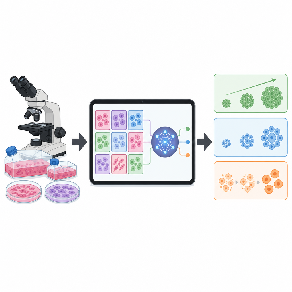
Teaching the computer to see like a cell expert
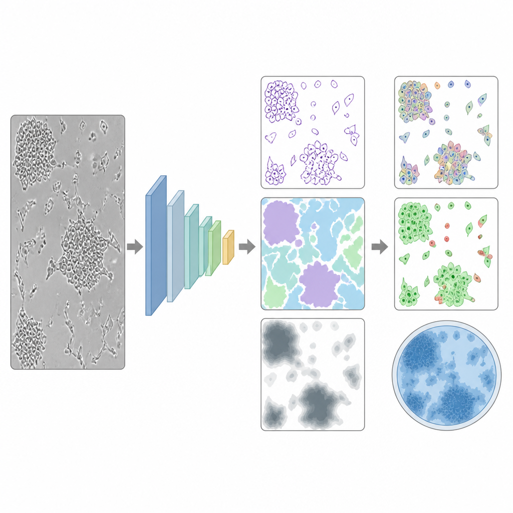
For judging how much of a surface is covered by cells, the team used a kind of image-analysis network that can turn each pixel into a simple “cell” or “not cell” decision. They iteratively trained and corrected this model until it reached more than 90 percent accuracy on independent test images. SnapCyte’s estimates of cell coverage were almost perfectly aligned with human experts and clearly surpassed several other popular software tools. The system also remained accurate when picture quality and resolution changed, suggesting that it can handle the uneven lighting, focus, and contrast that are typical in everyday lab work.
From coverage to counts and cell health
Counting individual cells and deciding which are alive is harder, especially when cells overlap or vary in size. To tackle this, the researchers adapted an existing cell-segmentation network and retrained it on images of many cell types, blood cells, and even plastic beads of known sizes. They used a “human-in-the-loop” process, repeatedly correcting errors and adding tougher examples until the model could find more than 95 percent of cells while rarely mistaking debris for cells. A companion model learned to estimate cell size and separate live from dead cells stained with common dyes. Across tests with mixed live and dead samples, SnapCyte’s results differed from expert human counts by less than five percent, while previous machine-learning approaches had much larger errors.
Putting AI to the test against classic lab methods
The team then asked whether SnapCyte could replace or match standard growth and toxicity tests used in research and drug development. They compared its measurements of cell coverage to direct cell counts, to color-based assays that track metabolism, and to a commercial live-cell imaging instrument. In multiple cell lines and drug-treatment experiments, SnapCyte’s readouts tracked closely with these established methods and often showed less variation from user to user. The tool could also estimate how long it takes cells to double in number and gave drug sensitivity values that matched published data while using fewer plates, fewer reagents, and much less hands-on time.
What this means for everyday lab work
Overall, the study shows that simple images of cell cultures contain enough visual information for a trained AI system to judge how many cells are present and how healthy they are, without touching the cells or adding chemicals. SnapCyte provides fast, consistent measurements that are largely independent of who is using it or what microscope they have. For working scientists, this could make routine checks on cell growth more reliable and less labor-intensive, while preserving the very same cells for later experiments.
Citation: Wong, C.P., Khazamipour, N., Aalibagi, S. et al. Enhanced precision in cell culture analytics: leveraging artificial intelligence for unbiased and non-destructive assessment of cell growth and viability. Cell Death Discov. 12, 234 (2026). https://doi.org/10.1038/s41420-026-03116-9
Keywords: cell culture, artificial intelligence, cell counting, cell viability, image analysis