Clear Sky Science · en
A comparative analysis of serum and tissue proteomic profiles in non-small cell lung cancer patients with or without brain metastasis
Why catching brain spread of lung cancer matters
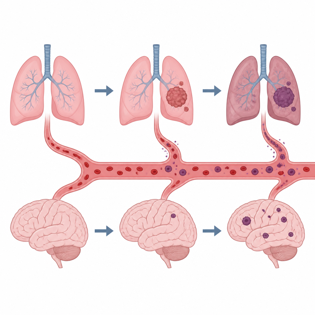
Non small cell lung cancer is common, and many patients eventually develop cancer spread to the brain. Once this happens, symptoms can worsen quickly and survival often shortens to just a few months. Today, doctors usually spot brain spread with MRI scans, which may miss the earliest changes and are not ideal for frequent, long term checks. This study asks a simple but important question: can a regular blood test reveal which lung cancer patients are at high risk of brain spread before serious damage occurs?
Looking for hidden clues in blood and tissue
To tackle this question, the researchers compared proteins in brain tumors that started from lung cancer with proteins in nearby normal brain tissue from the same patients. At the same time, they measured thousands of proteins in blood samples from people with lung cancer, lung cancer with brain spread, and healthy volunteers. Using a mass spectrometry method that can detect very low amounts of many proteins at once, they built one of the most detailed protein maps so far for this disease in both tissue and blood.
What changes when lung cancer reaches the brain
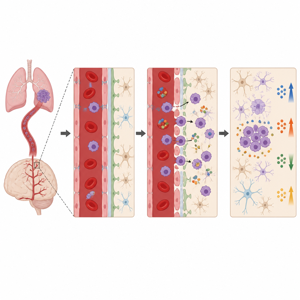
The team found that brain tumors from lung cancer look very different from nearby healthy brain at the protein level. Tumor tissue showed boosts in proteins involved in making and folding other proteins, stress responses inside cells, and processes that help cancer cells invade and remodel their surroundings. At the same time, proteins linked to normal brain growth, nerve connections, and synapses were reduced, reflecting how the tumor disrupts healthy brain function. In blood, they saw broad shifts in proteins related to immunity, transport, metabolism, and chemical signals called cytokines, which can help cancer cells cross into the brain and settle there.
Connecting tissue changes to a simple blood test
Because some proteins leak from tumors into the bloodstream, the researchers next looked for proteins that changed in the same direction in both brain tissue and blood. They identified a set of shared proteins involved in wound healing, protein maturation, and cell movement that were altered in patients with brain spread. Then they used machine learning, a type of computer based pattern finding, to sift through the blood data from lung cancer patients with and without brain metastases. By combining two different algorithms, they narrowed thousands of candidates down to a four protein panel that best separated the two groups.
Four proteins that flag higher brain risk
The final panel included four proteins: PSMA4, LAP3, LZIC, and RIC8B. In blood, three of these PSMA4, LAP3, and LZIC were consistently lower in patients whose lung cancer had already spread to the brain, compared with those whose cancer was still confined to the chest. When the authors tested this four protein set in statistical models, it correctly told apart patients with and without brain metastases in most cases, with high sensitivity and specificity. Follow up tests using standard lab kits and staining of tissue sections confirmed that especially PSMA4, LAP3, and LZIC also behave differently in brain metastases compared with primary brain tumors, suggesting they could help doctors tell these conditions apart under the microscope.
What this could mean for future care
For patients and clinicians, the main takeaway is that a carefully chosen combination of blood proteins can mirror what is happening inside brain metastases from lung cancer. While more work and larger, long term studies are needed before this becomes a routine test, the results show that a simple blood draw could one day help flag lung cancer patients at higher risk of brain spread, support earlier monitoring and treatment, and even aid in distinguishing brain metastases from primary brain tumors. In short, proteomics guided blood tests may move us closer to less invasive, more timely care for people facing this serious complication.
Citation: Zheng, Y., Xiong, Y., Ma, Y. et al. A comparative analysis of serum and tissue proteomic profiles in non-small cell lung cancer patients with or without brain metastasis. Cell Death Discov. 12, 230 (2026). https://doi.org/10.1038/s41420-026-03109-8
Keywords: non small cell lung cancer, brain metastasis, blood biomarkers, proteomics, early detection