Clear Sky Science · en
Low-dose TNF-α drives malignant progression and lipid metabolism in glioblastoma through the TRAF2-FASN axis
Why brain tumor fats matter
Glioblastoma is an aggressive brain cancer that is notoriously hard to treat and quick to return. This study looks at an unexpected helper that these tumors exploit: a common immune signal called TNF-α that, at low levels, can quietly push cancer cells to grow and stockpile fat. Understanding how this signal rewires tumor cells to hoard fats reveals a hidden vulnerability that might be turned into a new treatment target.
A mixed-message immune signal
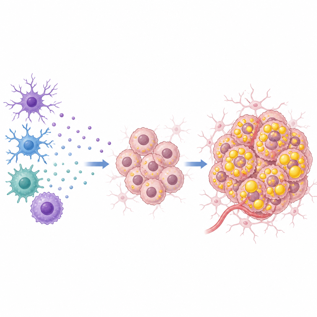
TNF-α is best known as a powerful alarm molecule of the immune system. At high doses, it can damage blood vessels feeding a tumor and trigger cancer cell death. Yet in many chronic diseases, including cancer, TNF-α lingers at much lower levels. The authors show that in glioblastoma, this low-dose state flips TNF-α from tumor enemy to tumor ally. Patient tumor samples and public cancer databases revealed that both main TNF-α receptors are unusually abundant in glioblastoma, and higher receptor levels are linked to shorter patient survival. This means glioblastoma cells are primed to “hear” even modest TNF-α signals in their surroundings.
Turning signals into fuel storage
In cell culture experiments, the team exposed glioblastoma cells to increasing doses of TNF-α and found a sweet spot around a low concentration where tumor cell growth peaked. At this dose, cells divided faster, migrated more readily, and invaded through barriers more aggressively. Gene activity profiles and microscopic staining pointed to a strong boost in fat handling: treated cells built up more lipid droplets and triglycerides, the storage form of fat. Blocking TNF-α receptors cut back this fat build-up, and patient tumors with more TNF-α showed richer deposits of lipid droplets, tying the lab findings to real human disease.
A protein relay that shields a fat-making engine
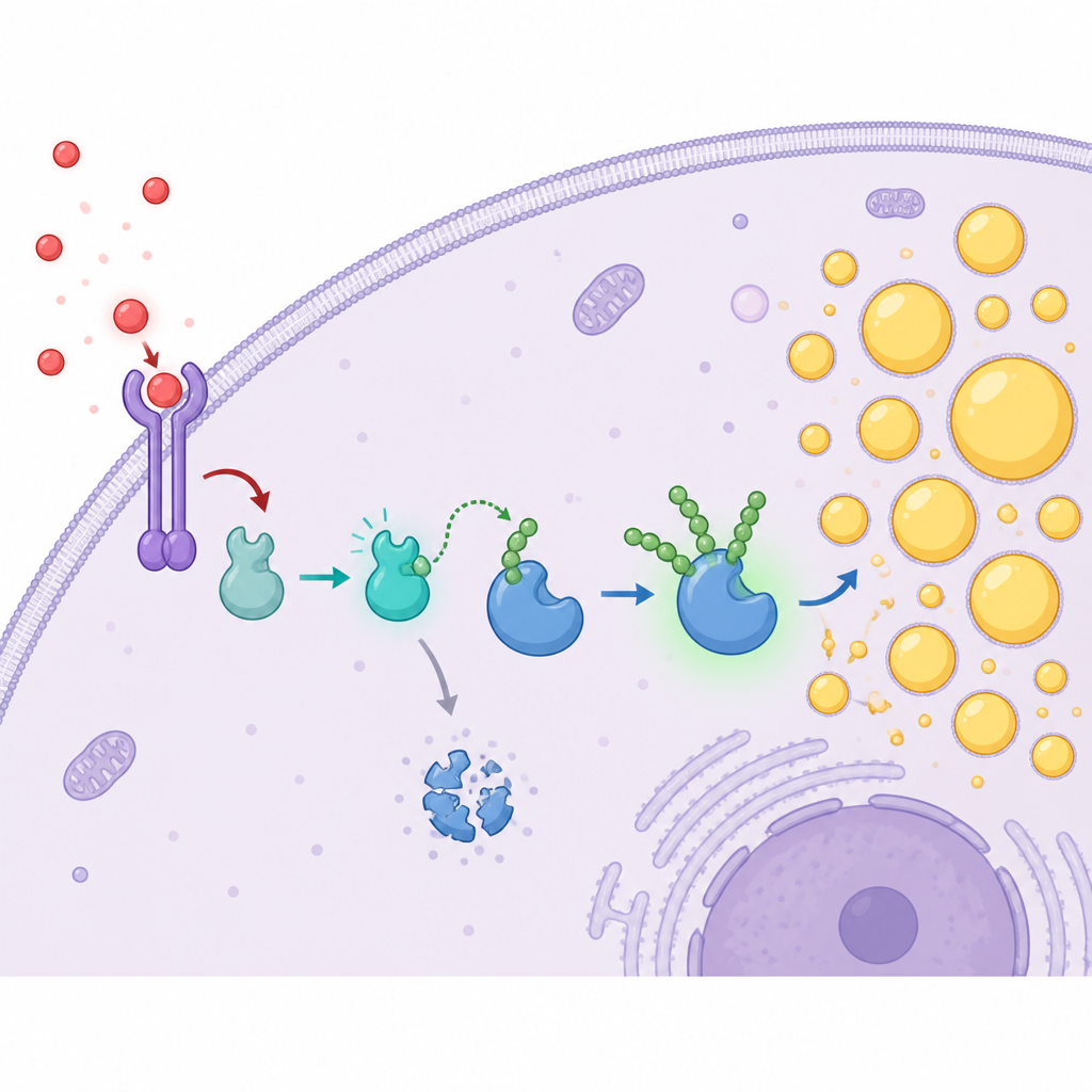
To uncover how TNF-α reshapes tumor fats, the researchers focused on a signaling adaptor protein called TRAF2 and a key fat-making enzyme named FASN. TRAF2 sits just downstream of TNF-α receptors and is often elevated in cancers. Here, TRAF2 levels were higher in glioblastoma cells and tumors than in normal brain and rose with tumor grade. Cells engineered to produce less TRAF2 grew more slowly, migrated and invaded less, and accumulated fewer lipid droplets and triglycerides, while extra TRAF2 had the opposite effects. Using protein interaction and imaging techniques, the team showed that TRAF2 physically binds FASN and makes FASN protein more abundant without changing its gene activity, hinting at a protective effect after the protein is made.
How protection from breakdown drives fat build-up
The study reveals that TRAF2 acts like a molecular bodyguard for FASN. TRAF2 carries a domain that functions as a tagging enzyme. It attaches special K63-linked ubiquitin chains onto FASN, a mark that stabilizes rather than destroys the enzyme. This modification slows FASN’s normal breakdown by the cell’s protein disposal machinery, extending its lifetime and allowing more fat to be made. Low-dose TNF-α strengthens the partnership between TRAF2 and FASN and increases these protective tags on both proteins. When TRAF2 was removed, TNF-α could no longer raise FASN levels or its ubiquitin tags, showing that TRAF2 is the crucial middleman turning an outside immune signal into extra fat production inside the tumor cell.
A plant-derived blocker with treatment potential
Armed with this detailed map, the team searched for small molecules that could interfere with TRAF2’s tagging activity. Through computer-guided screening, they pinpointed a compound called Jionoside B1 that binds to the TRAF2 region responsible for adding ubiquitin chains. In glioblastoma cells, Jionoside B1 lowered FASN protein levels, sped up FASN breakdown, reduced lipid droplets and triglycerides, and weakened cell growth, migration, and invasion. It also disrupted the TRAF2–FASN interaction and cut back the stabilizing ubiquitin chains on FASN. In mice with glioblastoma-like tumors in the brain, treatment with Jionoside B1 slowed tumor growth, supporting the idea that this pathway can be drugged in living organisms.
What this means for future brain cancer care
This work outlines a clear path by which low-dose TNF-α in the tumor microenvironment encourages glioblastoma cells to grow: it activates TRAF2, which then protects the fat-making enzyme FASN from breakdown, allowing tumor cells to stockpile fats needed for membranes, energy, and signaling. By exposing this TNF-α–TRAF2–FASN chain and showing that a small molecule can disrupt it, the study suggests a new angle for therapy that targets the metabolic lifelines of glioblastoma rather than the tumor DNA alone.
Citation: Cai, M., Liu, Y., Mao, X. et al. Low-dose TNF-α drives malignant progression and lipid metabolism in glioblastoma through the TRAF2-FASN axis. Cell Death Discov. 12, 242 (2026). https://doi.org/10.1038/s41420-026-03087-x
Keywords: glioblastoma, lipid metabolism, TNF-alpha, TRAF2 FASN axis, brain tumor signaling