Clear Sky Science · en
Brain metastases exhibit distinct spatial patterns of resident and infiltrating macrophages
Why brain spread of cancer matters
When cancers like lung, breast, or melanoma spread to the brain, they are hard to treat and often shorten life. This study looks not at the cancer cells themselves, but at the brain’s own helper cells that gather around tumors. By seeing where these cells sit in and around brain tumors, the researchers hope to guide future treatments that work with, or against, these helpers to better control disease.
The brain’s own cleanup crew
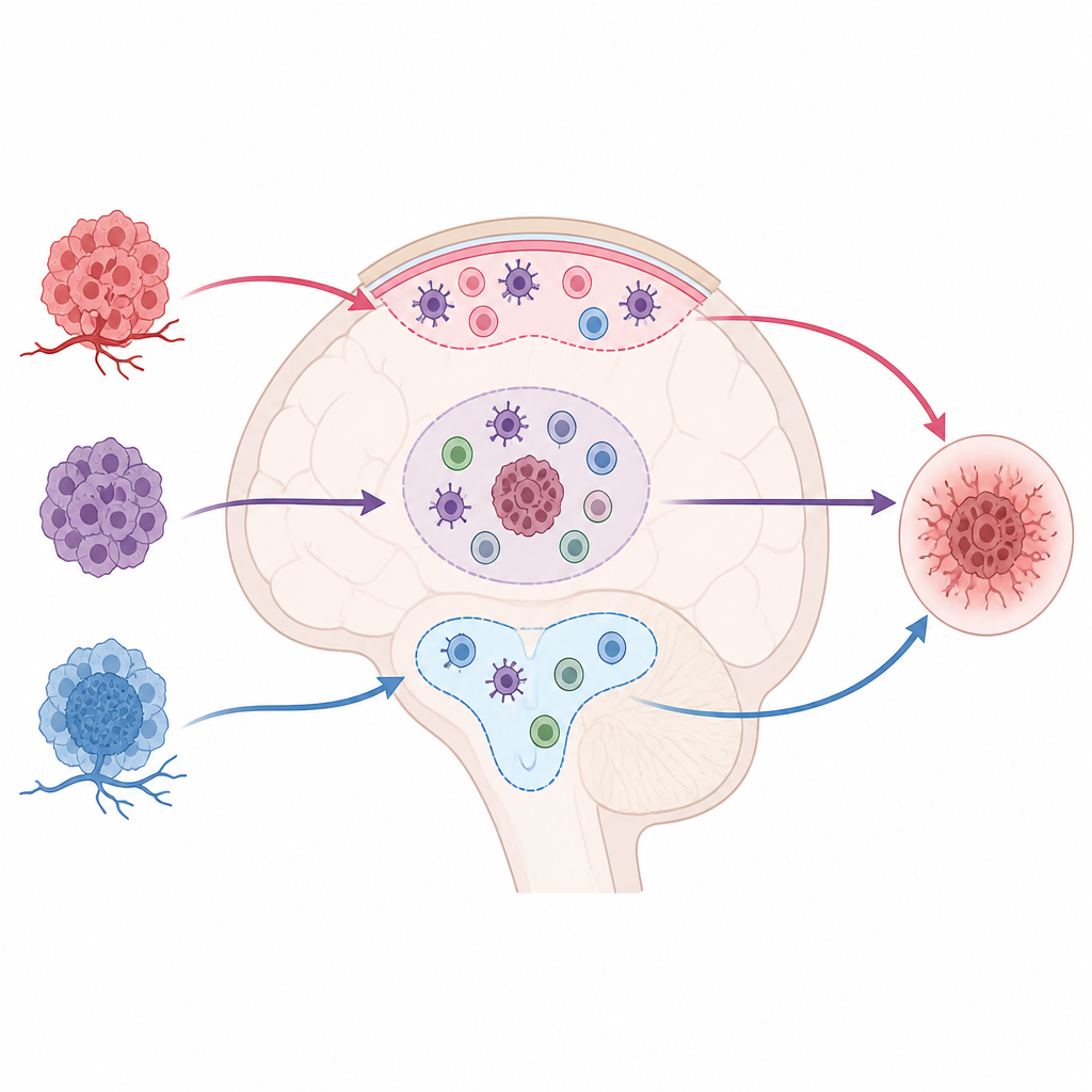
The brain is patrolled by several kinds of immune-like cells that act as a cleanup crew. Deep inside the brain live microglia, long-term residents that watch for damage or infection. Along the surfaces, coverings, and fluid-filled spaces sit border cells called border-associated macrophages, while another group of short-term visitors arrives from the blood as monocytes that turn into macrophages once inside. Together, these groups surround and enter brain tumors and are called tumor-associated macrophages. They can either help hold tumors in check or, in some cases, quietly support their growth.
Mapping who goes where in the brain
To learn how this cleanup crew behaves in different brain locations, the team used mouse models of three common cancers that spread to the brain: lung, breast, and melanoma. They implanted cancer cells so that tumors formed either in the brain tissue itself, in the fluid-filled ventricles, or along the delicate outer layers that wrap the brain. Using special genetic labels that light up specific cell lineages, they could tell apart long-term brain residents, border cells, and recent arrivals from the blood, then carefully counted and mapped these cells in and around tumors of different sizes.
Shifting roles as tumors grow
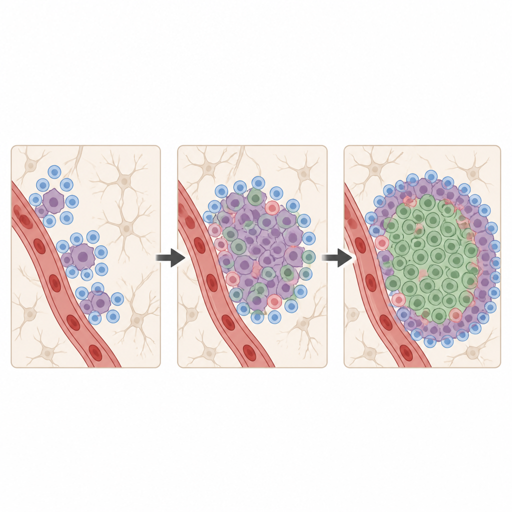
Inside the brain tissue, tiny early tumors were mostly surrounded by resident microglia. These cells multiplied locally and crowded around the tumor edge, with some slipping into the center as the growth enlarged. As tumors grew bigger and small spots merged into major masses, blood-borne macrophages became more and more common, particularly in the tumor core. This change in the balance between residents and newcomers depended on tumor size and also on which cancer type had seeded the brain, with breast cancer models drawing in especially large numbers of incoming cells.
Different brain niches, different defenders
The picture looked very different in tumors growing in the brain’s borders. In ventricular tumors that involved the choroid plexus, and in tumors along the thin coverings of the brain, microglia were largely absent. Instead, border-associated macrophages native to these sites expanded, while blood-derived macrophages infiltrated from nearby vessels. Melanoma metastases, especially those floating in the brain’s fluid spaces, showed strikingly fewer incoming macrophages than lung or breast cancer metastases. Across all sites, both resident and incoming cells showed signs of activation and a range of different states, suggesting a complex and varied immune environment.
What this means for future treatments
The study shows that there is no single, uniform immune setting around brain tumors. Rather, the mix of resident and incoming helper cells depends on where in the brain the tumor sits, how large it is, and which organ the cancer originally came from. For small tumors in brain tissue, resident microglia are the main responders, while in larger masses and in border regions, incoming macrophages and border cells play a growing role. For treatments that aim to alter or target these cells, it will not be enough to know the cancer type; doctors may also need to consider the exact brain compartment and stage of growth to choose the right strategy.
Citation: Ratzabi, A., Caspit, I.M., Telechi, I. et al. Brain metastases exhibit distinct spatial patterns of resident and infiltrating macrophages. Cell Death Discov. 12, 211 (2026). https://doi.org/10.1038/s41420-026-03084-0
Keywords: brain metastases, microglia, macrophages, tumor microenvironment, neuroinflammation