Clear Sky Science · en
Store-operated calcium entry drives alcohol-exacerbated neuroinflammation in retinal degeneration
When Drinking Meets Failing Eyes
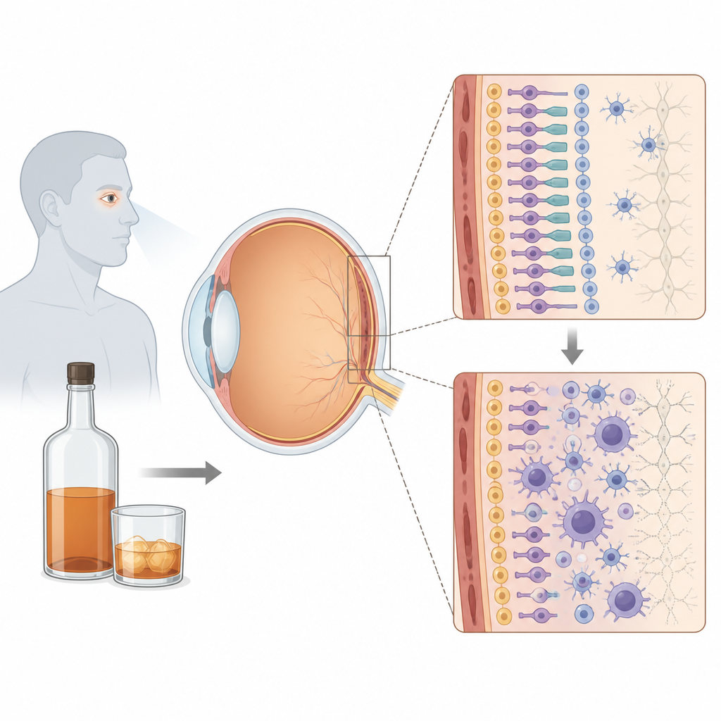
Retinitis pigmentosa is an inherited disease that slowly robs people of their sight. Many patients also face anxiety and depression, and some turn to alcohol to cope. This study asks a simple but important question: can alcohol make an already struggling retina even sicker, and if so, is there a way to tone down this damage by calming the brain’s immune cells?
The Brain’s Immune Cells in the Eye
The retina is not just a sheet of light-sensing nerve cells; it also contains support and immune cells that help keep it healthy. Among them are microglia, the brain’s resident immune sentinels, and Müller cells, long support cells that span the thickness of the retina. In diseases such as retinitis pigmentosa, microglia become activated and release inflammatory molecules, a process known as neuroinflammation. The authors focused on how these cells react when the retina is first stressed by oxidation and then exposed to alcohol, echoing what may happen in patients with eye disease who also drink heavily.
A Double Hit of Stress and Alcohol
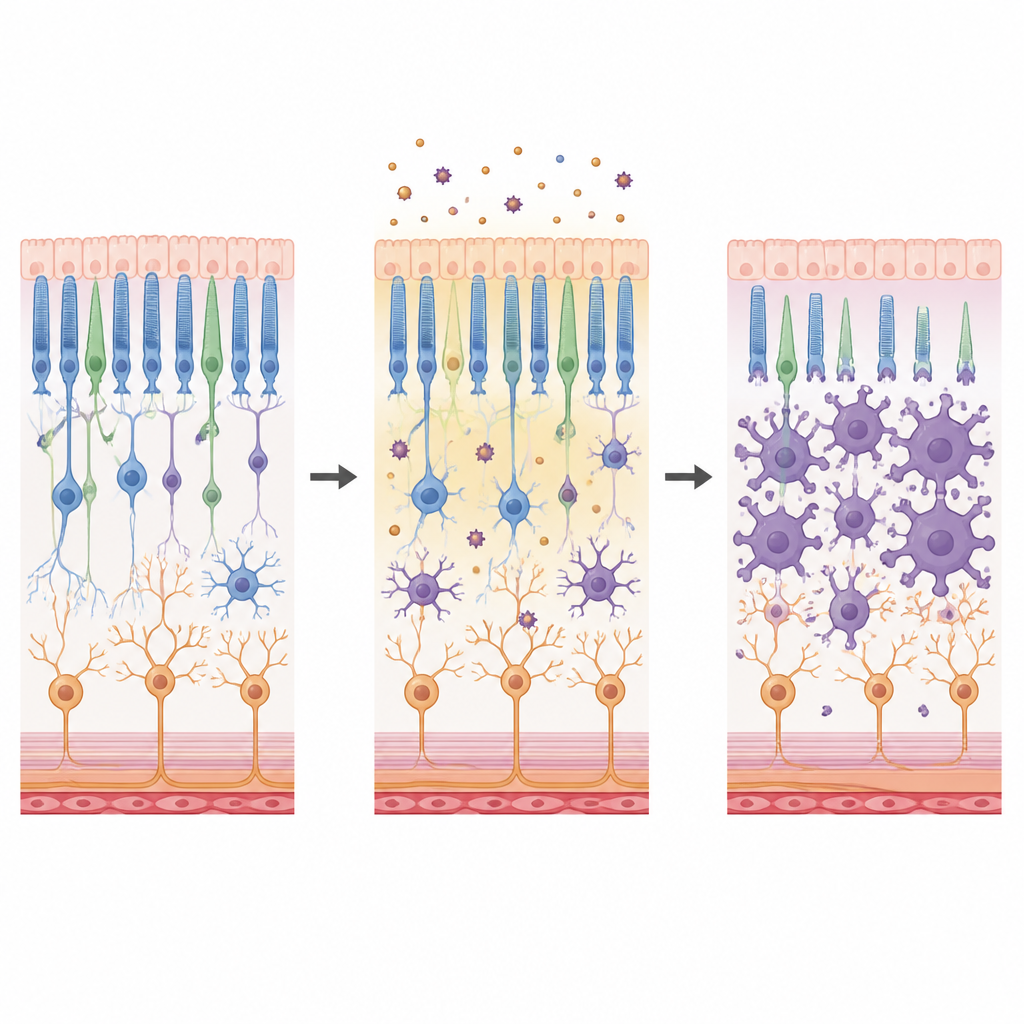
To mimic this “double hit,” the team grew mixed retinal cells in dishes and first exposed them to a mild dose of hydrogen peroxide to create oxidative stress. After this first insult, they added ethanol as a second blow. Under the microscope, microglia in this double-hit condition shifted from a delicate, branched shape to a rounder, amoeboid form linked with attack mode. At the same time, there was clear loss of light-sensing cells and other neurons, and their fine branches became shorter and fewer. Chemical measurements showed that inflammatory signals rose and formed tightly connected networks, painting a picture of a retina pushed into a more hostile state by alcohol on top of prior stress.
Turning Down the Calcium Signal
Microglia use calcium signals inside their cells as a control knob for activation. A major gateway for that calcium is a pathway called store-operated calcium entry, largely controlled by channels known as Orai. The researchers tested a drug called 2-APB, which can block this calcium flow. When they added 2-APB before the alcohol exposure in their cell cultures, microglia kept a more branched, less aggressive shape and shifted toward a less inflammatory state. Neurons survived better, with more of their normal long projections preserved. Further experiments using conditioned media showed that substances released from stressed microglia were especially toxic, and that blocking calcium entry in these cells, but not in Müller cells, reduced the harm to neighboring neurons.
What Happens in Diseased Eyes
The group then moved to a mouse model that naturally develops retinitis pigmentosa. They injected ethanol and 2-APB directly under the retina and examined the tissue a few days later. Even in this complex setting, 2-APB nudged microglia toward a more intricate, ramified appearance in both inner and outer retinal layers, suggesting a calmer, surveying state rather than an attack mode. Levels of several inflammatory messenger molecules, including IL-17, TNF-alpha, IL-10, and IL-13, dropped, and the network of interactions between these messengers became less tightly wired. Interestingly, simple surface markers that are often used to label “good” or “bad” microglia did not change, implying that shape and cytokine patterns are better indicators of their true behavior in this disease.
Limits and Hints for Future Care
Despite these changes in immune cells and inflammation, 2-APB did not stop the loss of light-sensing cells in the mouse retinas over the short time frame studied. The drug also has other targets besides the calcium channel of interest, so its benefits cannot yet be pinned to a single mechanism. Still, the work supports a clear idea for patients and clinicians: in a retina already under oxidative stress, alcohol can amplify damaging inflammation through calcium-driven activation of microglia. By dialing down this calcium signal, it may be possible to soften the inflammatory blow and better protect fragile retinal tissue from the added burden of alcohol and other systemic stresses.
Citation: Lima-Vasconcellos, T.H.d., Menezes, B.d.A., Móvio, M.I. et al. Store-operated calcium entry drives alcohol-exacerbated neuroinflammation in retinal degeneration. Cell Death Discov. 12, 222 (2026). https://doi.org/10.1038/s41420-026-03074-2
Keywords: retinitis pigmentosa, retinal inflammation, microglia, alcohol and vision, calcium signaling