Clear Sky Science · en
MAPK14/SLC7A11/GPX4 axis dysregulation drives podocyte ferroptosis via mediating glycerophospholipid metabolism
Why Kidney Cells Matter in Diabetes
People with long-standing diabetes often worry about their kidneys, because diabetic nephropathy is the leading cause of kidney failure worldwide. Yet we still lack medicines that can reliably halt the disease. This study digs deep into diabetic kidneys at single-cell and molecular levels to find out exactly which cells are in trouble, what is killing them, and how a natural compound from a traditional herb might protect them—while also uncovering simple urine molecules that could flag the disease early.
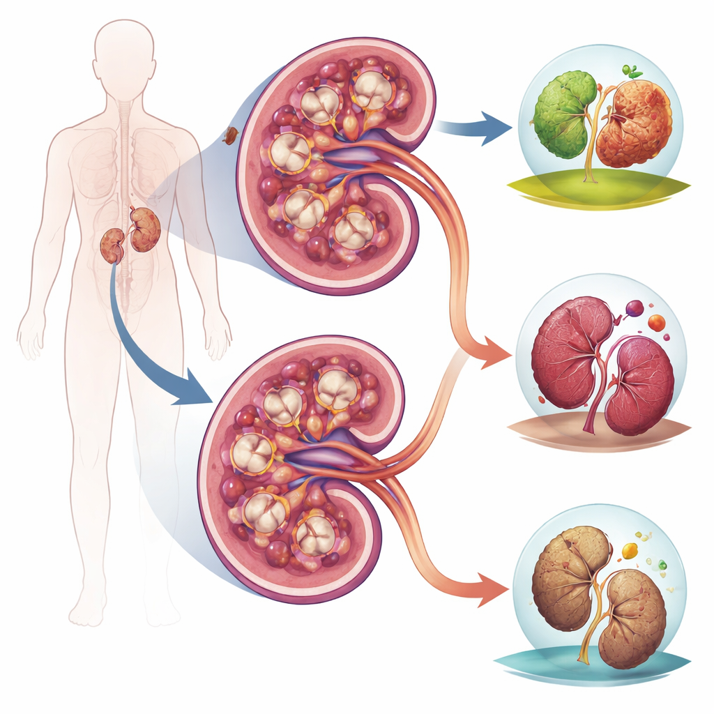
The Kidney’s Weak Link in Diabetes
The authors focused on podocytes, highly specialized cells that wrap around tiny blood vessels in the kidney’s filters and prevent protein from leaking into the urine. Using single-cell RNA sequencing on human kidney biopsies from people with diabetic nephropathy, they built a detailed “cell atlas” of the diseased organ. Among many kidney cell types, podocytes stood out as the central trouble spot: their gene activity patterns were the most altered by diabetes and were also the best at distinguishing diseased from healthy kidneys, suggesting these cells are both the main victims and strong candidates for early diagnosis.
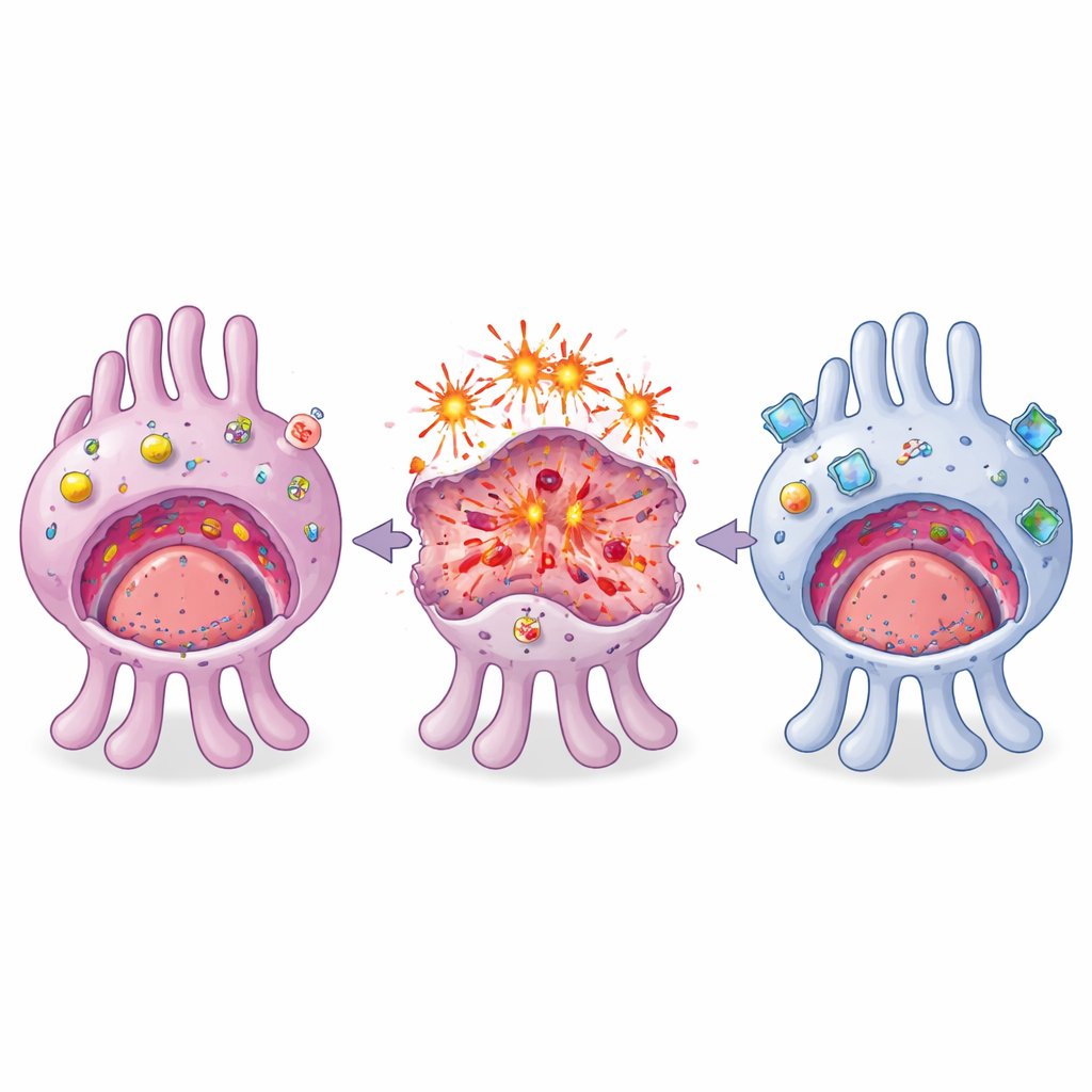
A New Form of Cell Death in Play
By zooming in on nearly two thousand individual podocytes, the team uncovered a specific type of regulated cell death called ferroptosis. Unlike classic cell suicide programs, ferroptosis depends on iron and runaway damage to fatty components of cell membranes. In diabetic podocytes, key genes that normally guard against this process—part of the MAPK14/SLC7A11/GPX4 axis—were misregulated. At the same time, genes involved in glycerophospholipid metabolism, which shapes the composition and vulnerability of membrane fats, were disturbed. Together, these changes create a perfect storm: membranes become loaded with easily oxidized fats while the cell’s defenses against lipid damage are weakened, making podocytes especially prone to ferroptosis.
Confirming the Culprit and Testing a Protector
To test whether this mechanism was conserved and druggable, the researchers turned to diabetic mice and cultured mouse podocytes. Single-cell sequencing in the mouse model revealed the same podocyte-centered metabolic disruptions seen in humans, supporting a core, cross-species mechanism. They then tested astragaloside IV (ASIV), a bioactive component from the herb Astragalus membranaceus, known for general kidney-protective effects. In diabetic mice, ASIV partially reset the overall kidney cell landscape toward a healthier pattern and, crucially, preserved podocyte numbers. At the molecular level, ASIV reversed diabetes-driven changes in the ferroptosis-related genes MAPK14, SLC7A11, and GPX4 and reduced biochemical signs of lipid damage, showing that its protective action centers on blocking ferroptosis rather than acting as a nonspecific antioxidant.
Mapping Metabolism in Kidney Tissue
The team next used spatial metabolomics, a technique that creates molecular images of tissue slices, to see where metabolic disruption occurs inside the kidney. They found that diabetic mice show sharply altered levels of several small molecules, especially in the kidney cortex where glomeruli and podocytes reside. Compounds linked to glycerophospholipid metabolism rose, while key antioxidants such as glutathione and its building block cystine fell—conditions that promote ferroptosis. ASIV treatment largely corrected these imbalances in both the cortex and the deeper medulla, indicating that the drug restores metabolic balance in specific anatomical regions, not just globally.
From Mechanism to Urine Tests
Finally, the researchers asked whether the same disrupted pathways leave a trace in patients’ urine. In a clinical metabolomics study comparing people with diabetic nephropathy to healthy volunteers, they found that a small set of metabolites—most notably serine, glutathione, and glycerol 3-phosphate—could distinguish patients with very high accuracy, rivaling or surpassing standard measures such as uric acid. Importantly, these molecules are directly tied to the ferroptosis and glycerophospholipid pathways uncovered in kidney cells, making them mechanistically meaningful biomarkers rather than vague markers of general illness.
What This Means for Patients
In everyday terms, this work shows that diabetic kidney damage centers on a specific kind of rust-like cell death in crucial filtering cells, driven by a mismatch between toxic fats and weakened antioxidant defenses. A natural compound, astragaloside IV, can interrupt this process in experimental models by stabilizing the key MAPK14/SLC7A11/GPX4 defense axis and normalizing kidney metabolism. At the same time, the same disturbed chemistry appears in urine, where simple metabolite measurements could one day allow earlier, more precise detection of diabetic kidney disease. Together, these findings sketch a complete path from microscopic mechanism to potential treatment and non-invasive testing.
Citation: Qiu, S., Xie, D., Guo, S. et al. MAPK14/SLC7A11/GPX4 axis dysregulation drives podocyte ferroptosis via mediating glycerophospholipid metabolism. Cell Death Discov. 12, 147 (2026). https://doi.org/10.1038/s41420-026-02990-7
Keywords: diabetic nephropathy, podocyte injury, ferroptosis, kidney metabolism, astragaloside IV