Clear Sky Science · en
WWP1 gain-of-function drives developmental anoikis through TGFβ pathway during neurodevelopment
When Brain Cells Lose Their Grip
Before birth, the brain is built from waves of newborn cells that must cling tightly to each other as they grow and migrate into position. This study explores what happens when a key molecular "traffic cop" called WWP1 becomes overactive. The researchers show that too much WWP1 makes young brain cells let go of their anchors, drift away, and die. Their work links a basic cell-cleanup system to early brain development and to severe childhood brain disorders.
Keeping Young Brain Cells Attached
In the developing brain, stem-like cells lining the fluid-filled ventricles divide and send newborn neurons outward to form the layered cerebral cortex. These progenitor cells rely on strong connections to their neighbors and to a belt-like surface at the ventricle wall to stay alive and organized. WWP1 is part of the cell’s protein-recycling machinery that normally fine-tunes many signaling pathways. The authors asked what would happen if WWP1’s activity was turned up too high during this delicate phase of brain construction.
Too Much WWP1, Cells Fall and Die
Using mouse embryos, the team artificially boosted WWP1 or introduced a known hyperactive version of the protein into developing cortex. Many of the labeled cells failed to reach their proper positions on time and showed clear signs of programmed cell death. The effect depended on WWP1’s enzymatic activity: when the catalytic site was disabled, the migration and survival problems disappeared. In human neural progenitor cells grown in the lab, extra-active WWP1 caused cells to round up, detach from the surface, float away, and then undergo caspase-driven death. This form of death after losing attachment, called anoikis, is well known in cancer biology but here appears in a developmental setting. 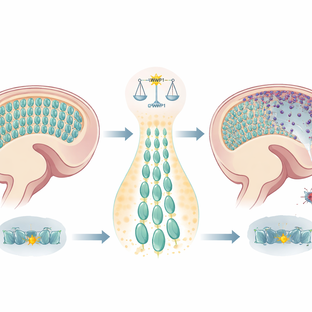
Broken Adhesion and Silent Survival Signals
Closer inspection showed that overactive WWP1 disrupted the actin-rich “belt” that normally holds progenitor cells at the ventricular surface. Many proteins that interact with WWP1 are involved in cell junctions, hinting that the ligase may be tagging adhesion components for destruction when it is overly active. The researchers also looked for signaling pathways that might counteract this damage. A screening experiment revealed that adding the growth factor TGFβ1 rescued many of the dying cells, both in culture and in mouse brains. Conversely, directly blocking the TGFβ pathway in otherwise normal embryos reproduced the same widespread detachment and death. Gene expression analysis in WWP1-hyperactive cells showed broad downshifts in genes tied to cell adhesion and TGFβ signaling, confirming that this survival pathway is dampened when WWP1 runs too hot.
From Molecules to a Child’s Brain Disorder
To connect these lab findings to human disease, the team studied a child with a severe developmental and epileptic brain condition marked by early-onset seizures and progressive brain atrophy. Genetic analysis uncovered a new, spontaneous change in the WWP1 gene that alters its catalytic domain. Structural modeling and biochemical tests showed that this variant makes WWP1 more flexible and more active, increasing its self-tagging behavior—a hallmark of gain-of-function. When this patient-derived variant was introduced into embryonic mouse brains, it triggered the same pattern of abnormal cell positioning and increased cell death as the known hyperactive WWP1 mutant. 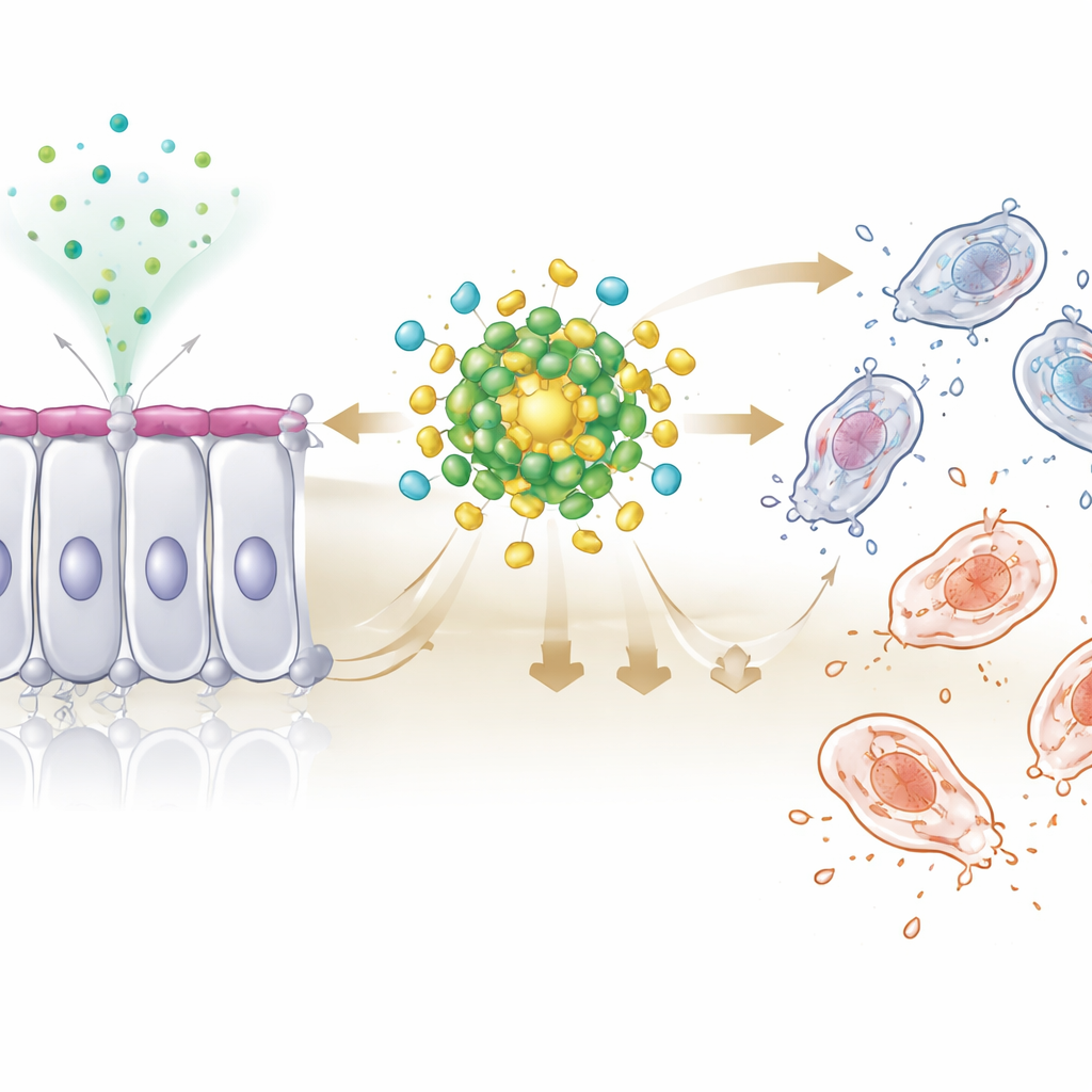
Why This Matters for Brain Health
Taken together, the study reveals that WWP1 must be kept in a narrow activity range to protect young brain cells. When WWP1 is overactive, it undermines the cell-to-cell bonds and survival signals that radial glia and newborn neurons depend on, causing them to detach and die instead of building healthy cortex. This “developmental anoikis” provides a plausible mechanism for certain neurodevelopmental disorders and epileptic syndromes linked to WWP1 and related genes. Because WWP1 and its pathways are already being explored as drug targets in cancer, this work opens the door to repurposing or refining such treatments to stabilize brain development in children who carry harmful WWP1 mutations.
Citation: So, K.H., Lee, S., Wong, J. et al. WWP1 gain-of-function drives developmental anoikis through TGFβ pathway during neurodevelopment. Cell Death Discov. 12, 133 (2026). https://doi.org/10.1038/s41420-026-02977-4
Keywords: neurodevelopment, cell adhesion, anoikis, TGF-beta signaling, WWP1