Clear Sky Science · en
Myeloid Mir34a suppresses initiation and progression of intestinal and colitis-induced colon cancers in APCmin mice
Why the body’s defenders matter in gut cancer
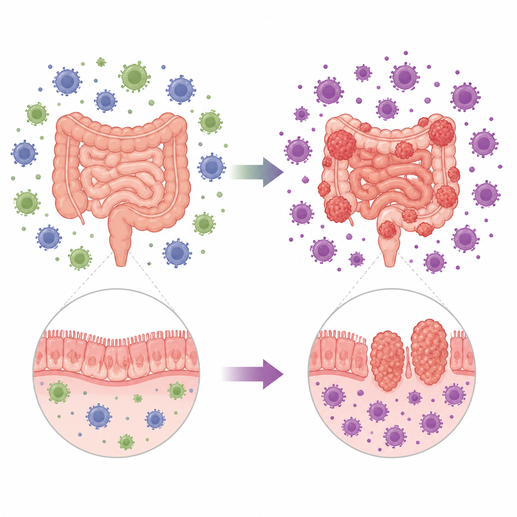
Colon and intestinal cancers do not grow in isolation; they develop within a busy neighborhood of immune cells. This study explores how one tiny genetic switch in certain immune cells can tip the balance between keeping early tumors in check and allowing them to grow, spread, and become life threatening in a common mouse model of intestinal and colon cancer.
A small genetic switch with a big impact
The researchers focused on a molecule called Mir34a, a short RNA controlled by the famous guardian gene p53. Instead of looking at Mir34a inside cancer cells, they studied it in myeloid cells, a family of immune cells that includes macrophages and neutrophils. Using mice that naturally develop many intestinal growths similar to early human colorectal tumors, they deleted Mir34a only in these myeloid cells. Mice lacking Mir34a in this compartment developed more intestinal tumors, and many of these growths advanced to aggressive, invasive cancers that are rarely seen in normal counterparts. The animals also died sooner, showing that this small genetic change in immune cells made the overall disease markedly worse.
How helper cells become enablers
Macrophages and neutrophils can either attack tumors or help them, depending on their “mood” or polarization. With Mir34a present, many tumor-associated macrophages and neutrophils in the gut showed a more inflammatory, tumor-fighting profile. When Mir34a was removed, these cells shifted toward a calmer, wound-healing mode that, in the context of cancer, actually supports tumor growth. Tumors from Mir34a-deficient mice were packed with macrophages and neutrophils, and chemical signals that call in and shape these cells were increased. The tumors contained more cells that divided rapidly and fewer that were undergoing cell death, indicating a more favorable environment for cancer expansion.
Ripple effects on other immune players
The change in myeloid cells also altered the balance of T cells, another key arm of the immune system. In mice lacking myeloid Mir34a, tumors contained more regulatory T cells, which are known to dampen immune responses, and fewer killer T cells equipped with enzymes for destroying abnormal cells. The team found higher levels of several known Mir34a target messages in tumors and in isolated macrophages, including factors that can boost invasion, shape macrophage behavior, and attract regulatory T cells. Together, these shifts created a more suppressive immune climate around the tumors, making it easier for cancer cells to grow, move, and eventually break through surrounding tissue.

From early changes to advanced disease
The influence of Mir34a was evident from the very beginning of tumor development. Young mice lacking Mir34a in myeloid cells showed more early abnormal structures in both small intestine and colon, and these early lesions were larger and more active. When the researchers triggered bowel inflammation with a chemical that models colitis, animals without myeloid Mir34a developed more colon tumors, including invasive cancers that penetrated deeply into the gut wall. In these inflamed tumors, macrophages and neutrophils again skewed toward tumor-helping states, and regulatory T cells accumulated, reinforcing an immune environment that favored progression rather than protection.
What this means for understanding gut cancer
This work shows that Mir34a in myeloid cells acts as a brake on intestinal and colon cancer by keeping local immune cells in a tumor-fighting state and limiting the buildup of suppressive T cells. When this brake is removed, the immune neighborhood around early lesions shifts to one that nurtures tumor initiation, growth, and invasion. While the study was done in mice, it supports the idea that restoring or mimicking this small RNA in specific immune cells could, in principle, help reprogram the tumor microenvironment in colorectal cancer toward stronger natural control of disease.
Citation: Chen, Y., Liu, F., König, J. et al. Myeloid Mir34a suppresses initiation and progression of intestinal and colitis-induced colon cancers in APCmin mice. Cell Death Dis 17, 458 (2026). https://doi.org/10.1038/s41419-026-08851-6
Keywords: colorectal cancer, tumor microenvironment, macrophages, microRNA, p53 pathway