Clear Sky Science · en
Neurochondrin drives colorectal cancer progression by modulating the PODXL–Ezrin axis and mitochondrial function
Why a brain protein matters in colon cancer
Colorectal cancer is one of the leading causes of cancer death, mainly because tumor cells can travel from the intestine to the liver. This study uncovers an unexpected player in that journey: neurochondrin, a protein best known for its role in the brain. The researchers show that when colorectal tumors make too much neurochondrin, the cancer cells gain extra energy, stick more tightly to other tissues, and are better able to spread to the liver. Understanding this hidden helper of metastasis could open new ways to slow or block advanced disease. 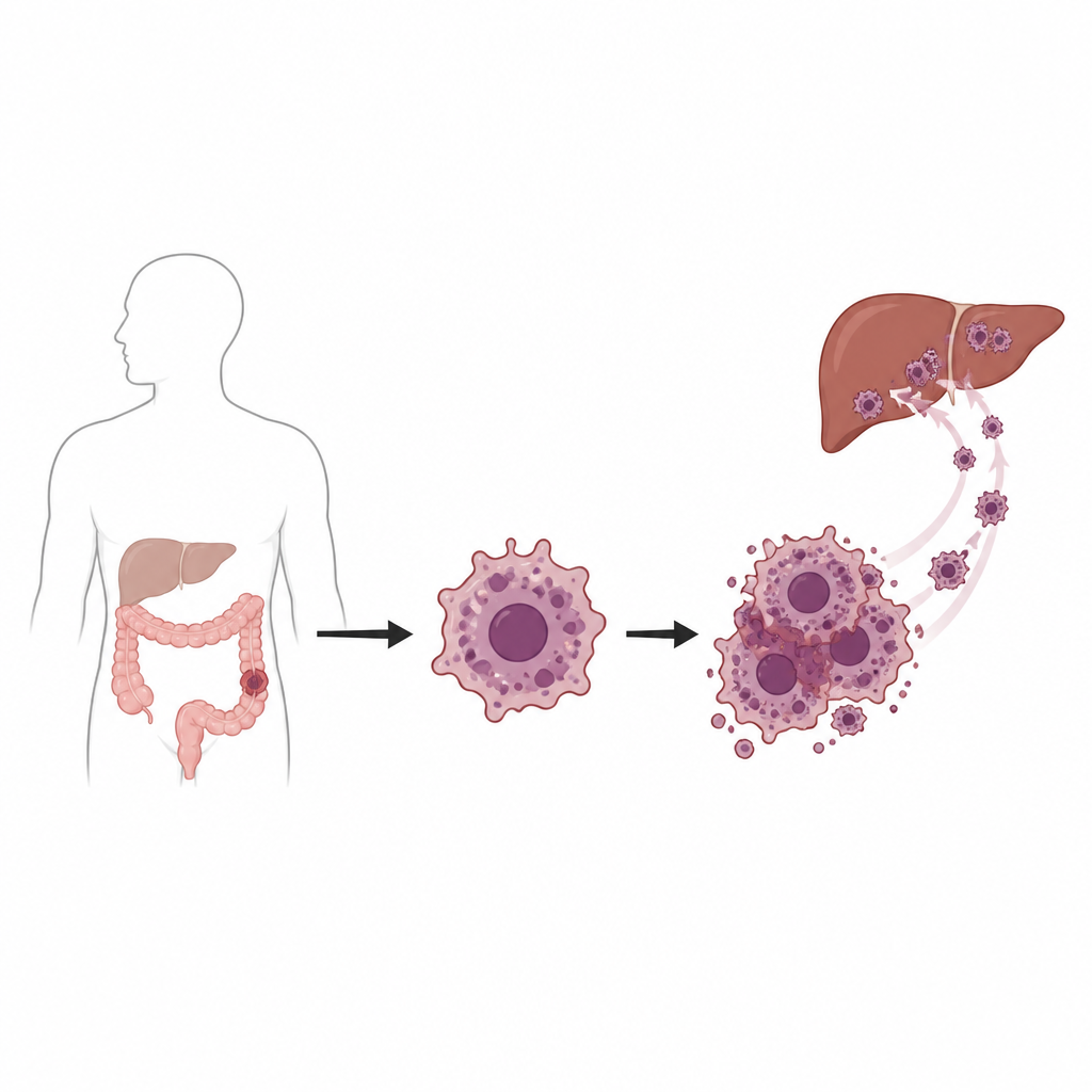
A hidden helper in deadly spread
Doctors have long known that liver metastasis is responsible for most deaths from colorectal cancer, but the detailed steps that allow tumor cells to colonize the liver remain only partly understood. Using pairs of closely related colon cancer cell lines that differ mainly in how easily they spread, the team noticed that the more aggressive cells consistently produced higher amounts of neurochondrin. They confirmed this pattern in many laboratory cell lines and in tumor samples from patients. Public cancer databases showed that tumors, especially advanced-stage ones, had more neurochondrin than nearby healthy tissue, and patients whose tumors expressed more of this protein tended to have shorter survival.
Putting neurochondrin to the test
To move from correlation to function, the scientists reduced neurochondrin levels in both weakly and strongly metastatic colon cancer cells. The altered cells grew more slowly, formed fewer colonies, and clung less well to a protein-rich surface. They were also more vulnerable to stress and cell death. When these neurochondrin-depleted cells were injected into mice, they struggled to form tumors under the skin, and far fewer reached or grew in the liver compared with control cells. Tumor size, growth rate, and the number of mice that actually developed liver metastases all shrank in line with how much neurochondrin had been removed, suggesting that the protein is important at several steps of tumor expansion and spread. 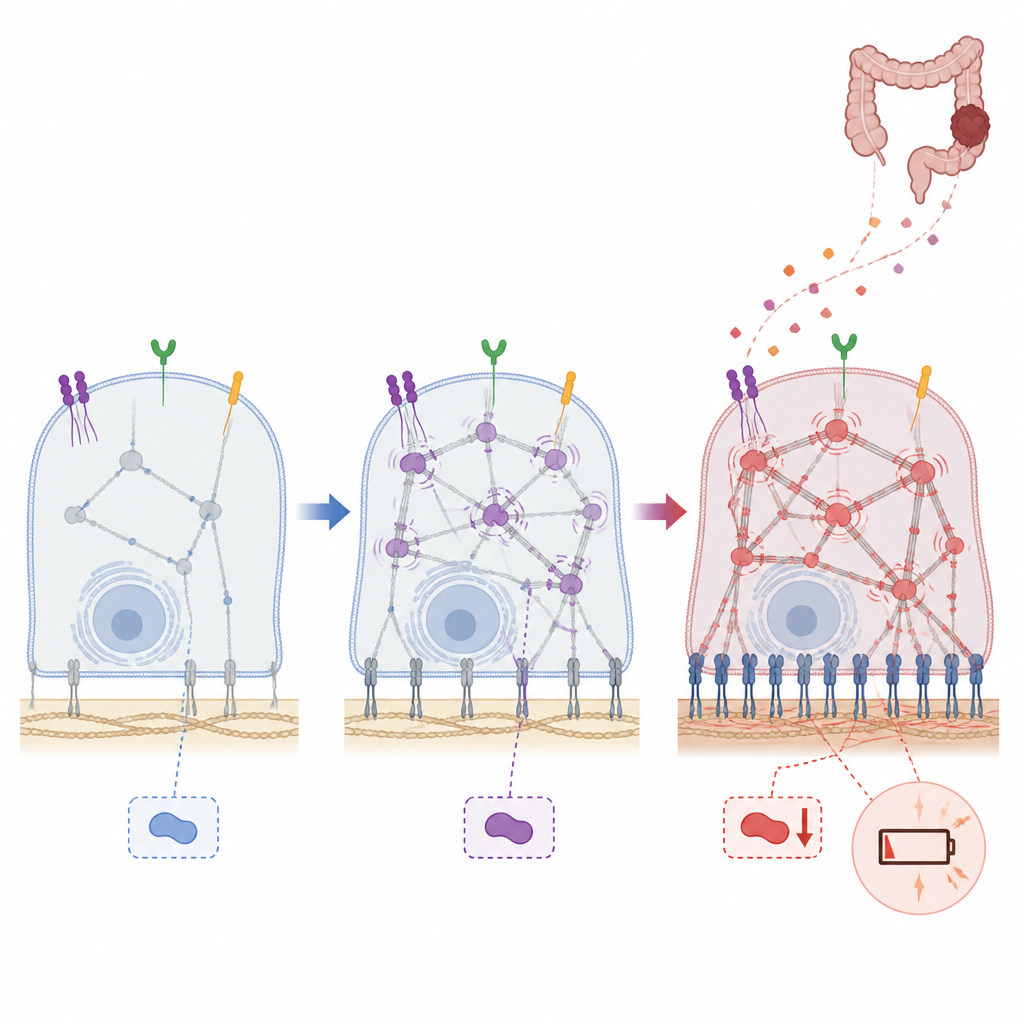
How stickiness and energy fuel metastasis
Diving deeper, the researchers mapped the proteins whose levels changed when neurochondrin was silenced. Many were involved in how cells attach, move, and communicate with their surroundings, as well as in how they handle energy within their mitochondria. One standout partner was podocalyxin, a surface molecule previously tied to poor outcomes and distant spread in several cancers. Podocalyxin connects to internal structural “linker” proteins such as ezrin and its relatives, which tie the cell’s outer membrane to its internal scaffolding and help control shape and movement. When neurochondrin was reduced, podocalyxin and ezrin family proteins dropped, signals from the growth receptor EGFR weakened, and cancer cells formed far fewer anchoring points called focal adhesions. In microfluidic channels lined with human blood vessel and connective tissue cells, neurochondrin-poor tumor cells had a much harder time attaching, hinting that they would be less able to exit the bloodstream and seed new sites.
Short-circuiting the cancer cell power supply
The protein surveys also pointed to changes in the tiny energy factories inside cells. In the most aggressive colon cancer cells, neurochondrin seemed to support robust mitochondrial activity and glycolysis, the pair of core processes that together power growth and movement. When neurochondrin was knocked down, oxygen consumption, spare energy capacity, and ATP production all fell, and the cells’ ability to ramp up sugar burning declined. Several mitochondrial components and antioxidant proteins went down, while some factors known to favor cell death went up. These shifts suggest that neurochondrin helps metastatic cells maintain the high, flexible energy output and stress resistance they need to survive in the bloodstream and adapt to new organs like the liver.
What this means for patients
Altogether, the work paints neurochondrin as a previously unrecognized organizer of both the “grip” and the “engine” of colorectal cancer cells. By boosting podocalyxin–ezrin signaling and mitochondrial function, it helps tumor cells attach to distant tissues and power their growth once they arrive. While much remains to be done before this knowledge reaches the clinic, neurochondrin and the network of proteins it controls now stand out as potential targets for therapies aimed at preventing or slowing the spread of colorectal cancer to the liver.
Citation: Garranzo-Asensio, M., Carral-Ibarra, E., Montero-Calle, A. et al. Neurochondrin drives colorectal cancer progression by modulating the PODXL–Ezrin axis and mitochondrial function. Cell Death Dis 17, 511 (2026). https://doi.org/10.1038/s41419-026-08747-5
Keywords: colorectal cancer, liver metastasis, neurochondrin, cancer cell adhesion, mitochondrial function