Clear Sky Science · en
UBC9-mediated SUMOylation of CORO1C drives lung adenocarcinoma progression via Arp2/3-dependent cytoskeletal remodeling
Why tiny cell switches matter in lung cancer
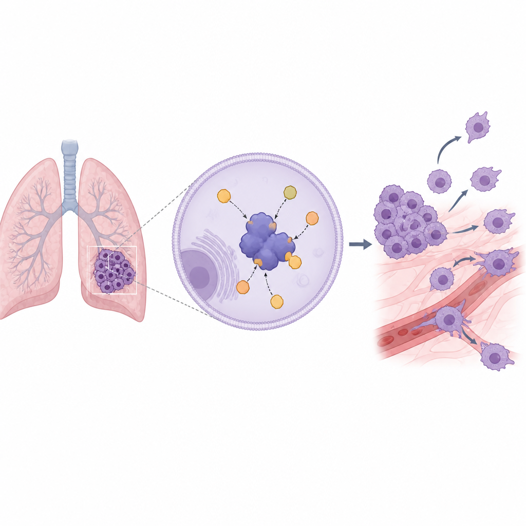
Lung adenocarcinoma is one of the most common and deadly forms of lung cancer, mainly because its cells are so good at growing, moving, and spreading. This study looks deep inside those cells to find out how a small molecular "switch" helps cancer cells reshape their internal skeleton, become more mobile, and form aggressive tumors. By understanding this switch, scientists hope to uncover new ways to slow or stop the spread of the disease.
A closer look inside lung tumor cells
The researchers began by asking whether a protein called UBC9, which helps attach small tags to other proteins, plays a special role in lung adenocarcinoma. Using public cancer databases and patient samples, they found that UBC9 is consistently higher in lung tumor tissues than in nearby healthy lung tissue. Patients whose tumors carried more UBC9 tended to have poorer survival, suggesting that this protein is not just present but linked to more aggressive disease. They also observed a general rise in tagged proteins in these tumors, hinting that this tagging process is unusually active in lung adenocarcinoma.
Switching off a driver of cancer behavior
To test whether UBC9 actually drives cancer behavior, the team used genetic tools to remove it from lung cancer cell lines grown in the lab. Without UBC9, these cells grew more slowly, formed far fewer colonies, and were much less able to move through artificial barriers meant to mimic tissue. When the altered cells were placed into mice, the resulting tumors grew more slowly, weighed less, and produced weaker signals in whole-body imaging. Taken together, these experiments show that UBC9 is not just a marker of bad outcomes; it actively supports the growth and spread of lung adenocarcinoma cells.
Finding the protein partner that shapes cell movement
Next, the researchers wanted to know which specific proteins UBC9 modifies to produce these effects. Using a fishing-like method that pulls down proteins bound to UBC9 and then identifies them by mass spectrometry, they pinpointed Coronin-1C, a protein that helps manage actin, the key building block of the cell’s internal skeleton. Coronin-1C normally works with a protein group called the Arp2/3 complex to build branched actin networks that push the cell’s edge forward, allowing it to crawl. The team showed that Coronin-1C receives small tags at three precise points, and that these tags depend on UBC9. When all three tag sites were removed, the modified version of Coronin-1C could no longer be tagged at all.
How cell skeleton changes fuel spread
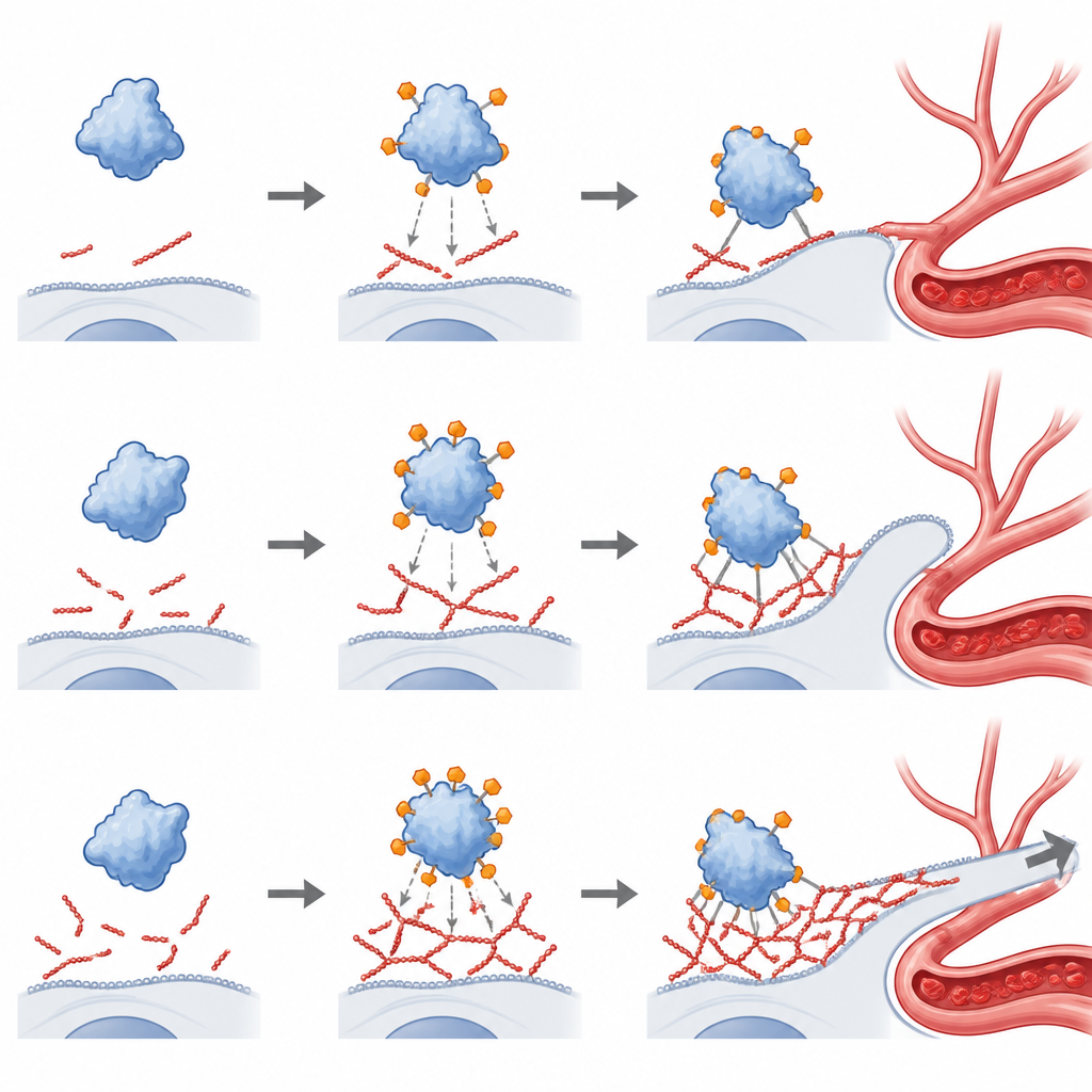
With that tagging system mapped out, the scientists asked what it actually does inside the cell. They compared normal Coronin-1C to a version that could not be tagged. Both versions sat in the same part of the cell, but only the normally tagged protein boosted cell growth, colony formation, and the ability to migrate and invade. In mice, cancer cells carrying the normal Coronin-1C formed larger tumors and many more metastatic spots in the lungs than cells carrying the tag-free version. Microscopic imaging revealed why: tagged Coronin-1C bound more tightly to the Arp2/3 complex and clustered with it at the leading edge of the cell, where dense actin branches formed powerful protrusions. The tag-free version showed weaker binding, poor overlap with Arp2/3, and a disorganized actin network.
Blocking the actin engine weakens cancer cells
To prove that this actin machinery is essential to the harmful effects of tagged Coronin-1C, the team used a chemical called CK-666 that holds the Arp2/3 complex in an inactive state. This treatment broke down the actin-rich protrusions normally driven by tagged Coronin-1C and sharply reduced cell growth, colony formation, migration, invasion, tumor size, and lung metastases in mice. In contrast, cancer cells with the tag-free Coronin-1C, which were already weakly aggressive, changed very little with the drug. These findings show that the UBC9–Coronin-1C axis pushes lung adenocarcinoma forward specifically by turning on Arp2/3-driven remodeling of the cell skeleton.
What this means for future treatments
In plain terms, this study reveals a chain of events: high levels of UBC9 in lung adenocarcinoma lead to extra tagging of Coronin-1C, which then grips more tightly onto the machinery that builds the cell’s inner scaffolding. That stronger grip allows cancer cells to reshape themselves, move more easily, and spread. Interrupting this chain, either at the level of UBC9, Coronin-1C tags, or the Arp2/3 complex, could offer new ways to make lung cancer cells less mobile and less likely to grow and metastasize. While much work remains before this can guide therapies, the study opens a clear new window into how tiny molecular switches control the deadly behavior of tumor cells.
Citation: Zhang, Z., Xiao, B., Jiang, Y. et al. UBC9-mediated SUMOylation of CORO1C drives lung adenocarcinoma progression via Arp2/3-dependent cytoskeletal remodeling. Cell Death Dis 17, 434 (2026). https://doi.org/10.1038/s41419-026-08653-w
Keywords: lung adenocarcinoma, cell cytoskeleton, protein modification, cancer cell migration, actin dynamics