Clear Sky Science · en
AURKA-mediated destabilization of SAPS3 drives ferroptosis evasion via 7-dehydrocholesterol biosynthesis in colorectal cancer
Why this research matters for cancer patients
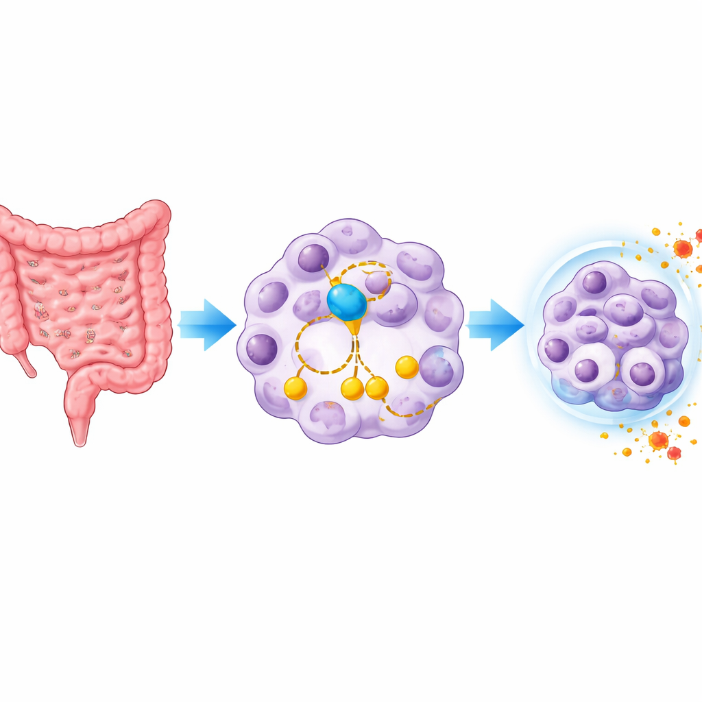
Many colorectal cancer patients eventually find that their tumors stop responding to chemotherapy. One emerging way to kill stubborn cancer cells is to trigger a form of iron‑driven, lipid‑based cell death called ferroptosis. Yet colorectal tumors are often strangely resistant to this process. This study uncovers how a well‑known cell division enzyme, Aurora kinase A (AURKA), rewires cholesterol‑related metabolism to help colorectal cancer cells dodge ferroptosis and chemotherapy – and shows that drugs blocking AURKA can make standard treatment work better.
An enzyme with a hidden survival trick
AURKA is best known for helping cells divide, and its overactivity is already linked to many cancers and poor outcomes. The authors started by mining large gene databases to find molecules tied both to colorectal cancer and ferroptosis. Among several candidates, shutting down AURKA made cancer cells grow more slowly and much more vulnerable to ferroptosis‑inducing compounds. Under the microscope, cells lacking AURKA showed shrunken, damaged mitochondria, high levels of reactive lipid molecules, and depleted antioxidant reserves – all hallmarks of ferroptosis. Analyses of tumor samples from patients confirmed that AURKA is markedly higher in colorectal cancers than in nearby normal gut tissue, and that patients with more AURKA tend to fare worse.
Cholesterol’s precursor as a shield against cell death
To understand how AURKA blocks ferroptosis, the team examined global gene activity when AURKA was removed. Surprisingly, many of the most strongly altered genes belonged to the cholesterol‑making pathway. One gene, DHCR7, stood out. It normally converts a molecule called 7‑dehydrocholesterol (7‑DHC) into cholesterol. Earlier work has shown that 7‑DHC can act as a powerful internal antioxidant that protects cell membranes from the damage that drives ferroptosis. In this study, loss of AURKA boosted DHCR7, which in turn drained 7‑DHC and lowered the cell’s ferroptosis resistance. Blocking DHCR7 genetically or with a drug restored 7‑DHC levels and made cells resistant to ferroptosis again, while adding extra 7‑DHC alone was enough to protect AURKA‑deficient cells. These results reveal that AURKA steers whether 7‑DHC is kept as a shield or converted away.
A molecular relay connecting metabolism and cell survival
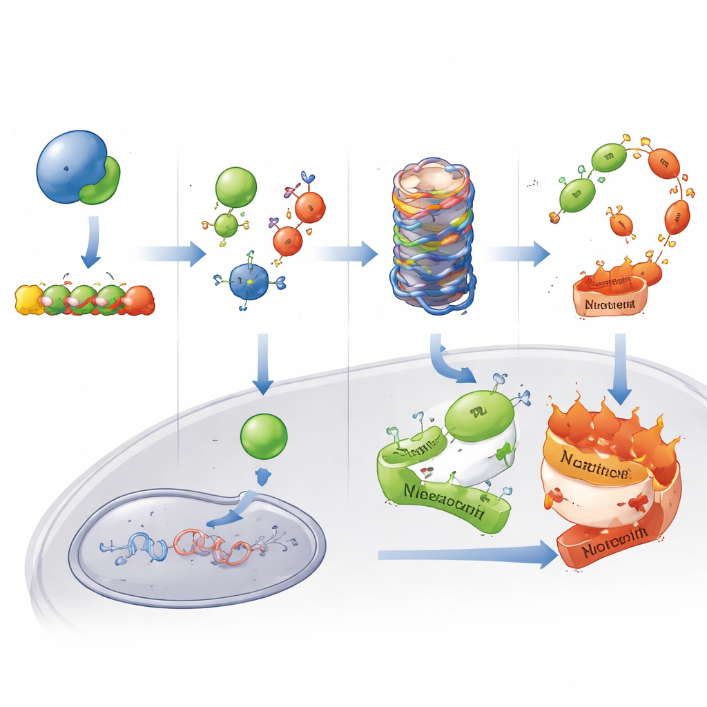
The researchers then traced the upstream signaling that links AURKA to DHCR7. They found that AURKA physically binds to a regulatory protein called SAPS3, which normally helps the enzyme PP6 keep the energy sensor AMPK in check. AURKA tags SAPS3 with phosphate groups at two specific sites, marking it for destruction by the cell’s protein disposal machinery. When AURKA is abundant, SAPS3 levels stay low, AMPK remains more active, and this dampens the movement of the cholesterol‑regulating factor SREBP2 into the nucleus. As a result, DHCR7 is kept down and 7‑DHC accumulates, buffering lipids against ferroptotic damage. When AURKA is removed or pharmacologically inhibited, SAPS3 stabilizes, AMPK activity drops, SREBP2 enters the nucleus, and DHCR7 turns on, tipping the balance away from 7‑DHC and toward ferroptosis sensitivity.
Turning a resistance mechanism into a therapeutic target
Because ferroptosis can complement standard chemotherapy, the team asked whether disabling AURKA might make common drugs like 5‑fluorouracil and oxaliplatin more effective. In cell cultures and mouse tumor models, genetic loss of AURKA or treatment with the AURKA inhibitor alisertib significantly boosted the tumor‑killing impact of chemotherapy. Tumors shrank more, showed higher lipid damage, and displayed more signs of both ferroptosis and apoptosis (a different form of cell death). In patient‑derived tumor grafts grown in mice, the combination of alisertib and 5‑fluorouracil was clearly superior to either agent alone. Finally, in clinical samples from colorectal cancer patients treated with chemotherapy, those whose tumors expressed more AURKA had shorter times before the disease progressed and poorer overall survival, supporting AURKA as a marker of chemoresistance.
What this means for future colorectal cancer treatment
This work reveals an unexpected link between a cell division kinase, cholesterol‑related metabolism, and resistance to lipid‑based cell death in colorectal cancer. By destabilizing SAPS3 and tuning the AMPK–SREBP2–DHCR7 pathway, AURKA preserves 7‑DHC and helps cancer cells survive both ferroptosis inducers and standard chemotherapy. In practical terms, the findings suggest that testing tumors for AURKA levels could help predict who will respond poorly to chemotherapy, and that pairing AURKA inhibitors such as alisertib with existing drugs might unlock a more powerful, ferroptosis‑driven attack on otherwise resistant colorectal cancers.
Citation: Gao, J., Zhang, W., Chen, L. et al. AURKA-mediated destabilization of SAPS3 drives ferroptosis evasion via 7-dehydrocholesterol biosynthesis in colorectal cancer. Cell Death Dis 17, 361 (2026). https://doi.org/10.1038/s41419-026-08549-9
Keywords: colorectal cancer, ferroptosis, cholesterol metabolism, Aurora kinase A, chemoresistance