Clear Sky Science · en
GABAergic ventrolateral preoptic projection to dorsomedial hypothalamus recapitulates post-ischemic neuroprotection by hypothermia
Cooling the Brain from the Inside
After a stroke, cooling the brain can dramatically limit damage, but current medical cooling methods often cause dangerous side effects such as heart rhythm problems and infections. This study in mice uncovers a built‑in brain circuit that can gently lower body temperature from within, protecting brain tissue after stroke without physically chilling the patient. Understanding this internal “thermostat switch” could open the door to safer treatments that harness hypothermia’s benefits while avoiding its risks.
The Problem with External Cooling
Doctors have long known that reducing body temperature helps injured brain cells survive after events like stroke or cardiac arrest. Cooling slows metabolism, stabilizes blood flow, and dampens inflammation. Yet large clinical trials in stroke patients have been disappointing: external cooling through cold blankets, ice packs, or chilled blood has not improved recovery overall. The main reason is that intense cooling stresses the rest of the body, disrupting heart rhythm, blood clotting, and immune defenses. The authors therefore asked a different question: instead of forcing the body from the outside, could they tap into the brain’s own temperature control centers to produce a milder, safer cooling that still protects the brain?
A Hidden Switch Between Two Brain Hubs
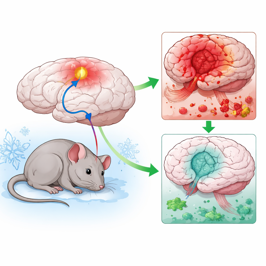
The team focused on two tiny regions deep in the hypothalamus, a brain area that regulates temperature. One region, the ventrolateral preoptic area, and another, the dorsomedial hypothalamus, are linked by nerve fibers that release the calming messenger GABA. Earlier work suggested that this connection acts as a brake on heat production: when the preoptic area activates, it quiets cells in the dorsomedial hypothalamus, leading to a drop in body temperature. Using advanced genetic tools in specially bred mice, the researchers could either silence the dorsomedial cells directly or turn on the preoptic‑to‑dorsomedial pathway with light. In both cases, mice became mildly hypothermic by about 2°C, moved around less, and showed no obvious abnormal behavior—consistent with a controlled, natural cooling response rather than a state of illness.
Less Blood Rush, Less Reperfusion Injury
The key test was whether this brain‑driven cooling would shield the brain during and after an experimental stroke, created by briefly blocking a major artery. When the scientists silenced the dorsomedial neurons before the blockage, or activated the preoptic pathway, the resulting brain lesions were smaller, swelling was reduced, and neurological scores improved. Imaging of blood flow revealed that, in cooled mice, the surge of blood back into the brain after the artery reopened was blunted and rose only slowly. This is important because an abrupt rush of warm, oxygen‑rich blood can paradoxically worsen damage—a process called reperfusion injury. By slightly constricting blood vessels and lowering metabolic demand, the hypothermic state appeared to cushion the brain against this secondary wave of harm, as shown by fewer cells with fragmented DNA, a sign of irreversible injury.
Keeping Support Cells in a Helpful Mode
Beyond neurons, the study examined astrocytes, star‑shaped support cells that can either help or hurt surrounding nerve cells after stroke. In untreated mice, astrocytes became strongly reactive and shifted into a “toxic” metabolic state, marked by high levels of a protein called PKM2 that pushes them toward harmful behavior. In animals cooled through the hypothalamic circuit, astrocytes remained closer to their normal, home‑supporting role: their activation marker decreased, PKM2 levels fell in both astrocytes and other cells, and survival of neurons in vulnerable regions improved over the following days. These findings suggest that engaging the internal cooling pathway does more than lower temperature—it also stabilizes the local environment that neurons depend on.
Toward Safer Brain Cooling Therapies
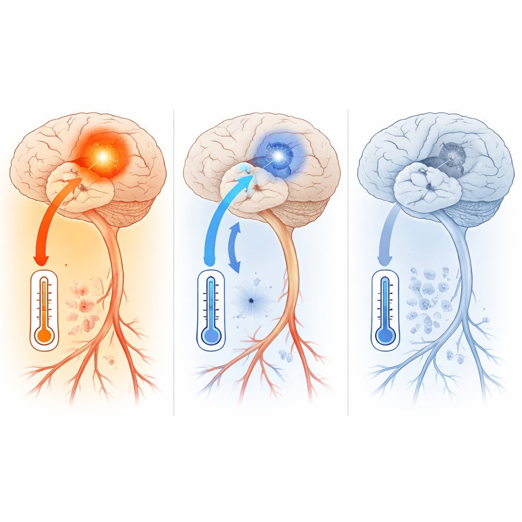
In plain terms, this work shows that flipping a specific “cooling switch” inside the brain can mimic the protective effects of traditional hypothermia while potentially sidestepping many whole‑body side effects. By dialing down a heat‑promoting hub in the hypothalamus, mice experienced gentle cooling, reduced blood‑flow surges, calmer support cells, and smaller strokes. Although the current tools—genetic switches and implanted optical fibers—are not ready for patients, the same pathway might one day be reached noninvasively through advanced brain stimulation. If so, future stroke treatments could activate the brain’s own thermostat to protect vulnerable tissue, offering a new route to improve recovery without putting the rest of the body at risk.
Citation: Dilsiz, P., Ozpinar, A., Balaban, B. et al. GABAergic ventrolateral preoptic projection to dorsomedial hypothalamus recapitulates post-ischemic neuroprotection by hypothermia. Cell Death Dis 17, 304 (2026). https://doi.org/10.1038/s41419-026-08536-0
Keywords: stroke, therapeutic hypothermia, hypothalamus, neuroprotection, astrocytes