Clear Sky Science · en
Evaluation of non-canonical p53 functions in DNA replication and recombination for variant classification
Why this research matters for families with cancer risk
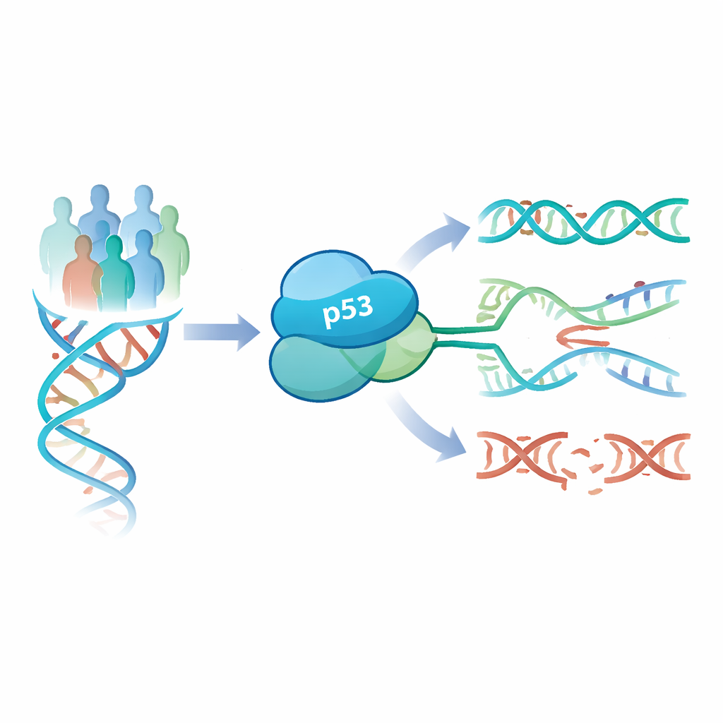
Many families affected by breast and ovarian cancer receive genetic test results that are difficult to interpret, especially when changes are found in a key cancer-protection gene called TP53. Some changes are clearly dangerous, some clearly harmless, but many fall into a gray zone called “variants of unknown significance.” This study explores a new way to tell which TP53 changes truly raise cancer risk by looking at how they influence the copying and repair of DNA, potentially helping doctors give clearer answers to patients.
A guardian gene with more than one job
TP53 encodes the p53 protein, often called the “guardian of the genome” because it helps prevent cells from turning cancerous. For years, most tests of TP53 variants focused on one main role of p53: turning other genes on and off to slow cell growth or trigger cell death after damage. These “canonical” functions have been widely used to classify variants as harmful or harmless. However, newer research shows that p53 also works directly at the DNA itself, especially when the cell’s copying machinery runs into obstacles. These more recently recognized “non-canonical” roles—such as controlling how DNA is replicated and repaired—might reveal problems that standard tests miss.
Following DNA repair instead of just gene switches
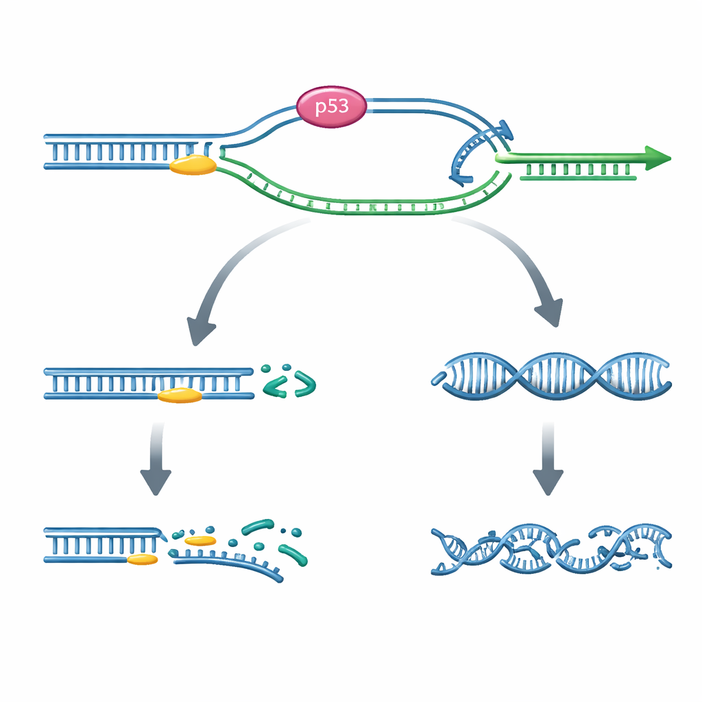
The authors studied 23 uncertain TP53 variants, along with 20 variants already labeled as clearly benign or clearly pathogenic, all identified in German families tested for hereditary breast and ovarian cancer. They used two cell-based tests that focus on what happens when DNA replication faces roadblocks. In the first test, cells carry a special DNA reporter that only lights up when a broken or blocked DNA stretch is successfully bypassed using a precise repair process called recombination. By introducing different TP53 variants into these cells and counting how often recombination occurred, the researchers could measure how well each version of p53 supported this “safe detour” around replication barriers.
What the recombination test revealed
The recombination assay clearly separated the known harmless and harmful variants: benign or likely benign TP53 variants consistently showed high levels of recombination, while pathogenic or likely pathogenic variants showed low levels. When the uncertain variants were tested, about one-third fell into the same activity range as the benign group, and another third into the clearly defective, pathogenic-like range. This means that eight previously unclear variants now emerge as strong candidates for reclassification. Importantly, the recombination scores lined up well with results from four large earlier studies that examined traditional p53 functions like gene activation and cell survival, reinforcing that this new assay is both reliable and informative.
Why measuring replication speed is not enough
In a second approach, the team used a DNA “fiber” assay that directly measures how fast new DNA is made along individual replication tracks. While earlier work showed that normal p53 slows replication slightly to allow safe repair, the pattern here was more complicated. Some clearly harmful variants did speed up replication as expected, but others did not, and several benign variants overlapped with the pathogenic range. Overall, this test did not cleanly distinguish harmful from harmless TP53 variants. The authors suggest that replication speed is influenced by many overlapping processes and is technically harder to measure robustly, making it less useful as a standalone classifier.
From protein structure to cancer risk
To understand why some variants affected recombination more than traditional p53 activities, the researchers also modeled the three-dimensional structure of the altered proteins. They found that certain “separation-of-function” variants subtly changed flexible surface loops or the way p53 assembles into its four-part, working form. Some of these changes appeared to spare basic gene-control functions while weakening p53’s ability to guide safe recombination at stalled replication forks. Intriguingly, the authors point out that such selective loss of recombination function may be particularly important in breast cancer, suggesting that not all harmful TP53 variants act through the same pathways.
What this means for patients and clinicians
By tracking how different TP53 variants influence a specific, non-canonical repair pathway, this study demonstrates that a recombination-based assay can sharply distinguish between low-risk and high-risk variants, including those with only subtle protein changes. While current international guidelines still rely heavily on older functional tests, the authors argue that adding recombination measurements could greatly improve the classification of borderline or low-penetrance TP53 variants in hereditary breast and ovarian cancer. For patients and families, this could translate into clearer risk estimates and more tailored surveillance or prevention strategies in the future.
Citation: Jansche, R., Heitmeir, B., Faust, U. et al. Evaluation of non-canonical p53 functions in DNA replication and recombination for variant classification. Cell Death Dis 17, 292 (2026). https://doi.org/10.1038/s41419-026-08463-0
Keywords: TP53 variants, p53 DNA repair, breast cancer risk, genetic variant classification, homologous recombination