Clear Sky Science · en
A tissue engineering approach to regenerate the cranial suture skeletal stem cell niche with a multicompartment biomaterial scaffold
Why soft seams in the skull matter
The bones of a baby’s skull are separated by soft seams called sutures, which leave room for the growing brain. In a condition known as craniosynostosis, some of these seams close too early, forcing the skull to grow in abnormal ways and sometimes putting pressure on the brain. Today the only real treatment is major surgery to cut and reshape bone. This study explores a very different idea: using a smart, sponge-like material to rebuild the skull’s natural growth seam so the head can continue to grow more normally.
A new way to think about skull surgery
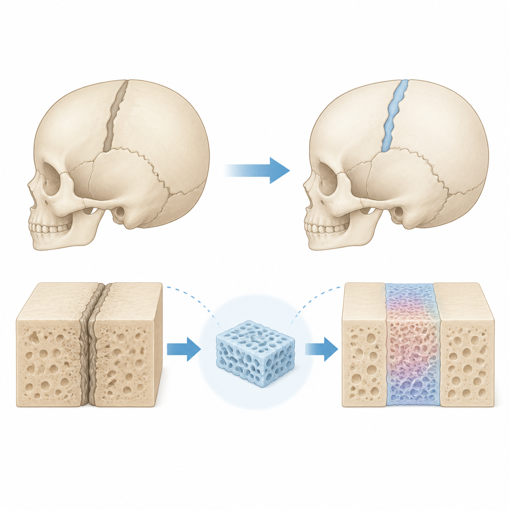
Standard operations for craniosynostosis remove fused bone but do not replace the missing living seam. As a result, the gap often fills back in with solid bone, and children may need more surgeries. The authors suggest a tissue engineering approach instead. Rather than only cutting bone, surgeons would also implant a custom scaffold that recreates the environment where the skull’s own stem cells usually live. These skeletal stem cells normally sit in the suture and generate new bone in a controlled way as the brain grows. When they are lost or forced to become bone too quickly, the seam fuses.
Designing a smart three-part scaffold
The team built a “triphasic” scaffold from a medical-grade biodegradable plastic. Its key feature is a patterned system of pores: a narrow central zone made of very small pores, sandwiched between two zones with much larger pores. In earlier work, the researchers had shown that small pores help stem cells stay in a more primitive, flexible state, while large pores encourage them to become bone and support blood vessels. Using a sugar template and a layer-by-layer process, they created a cylinder with sharply defined regions that allow cells to move through but still maintain distinct microenvironments.
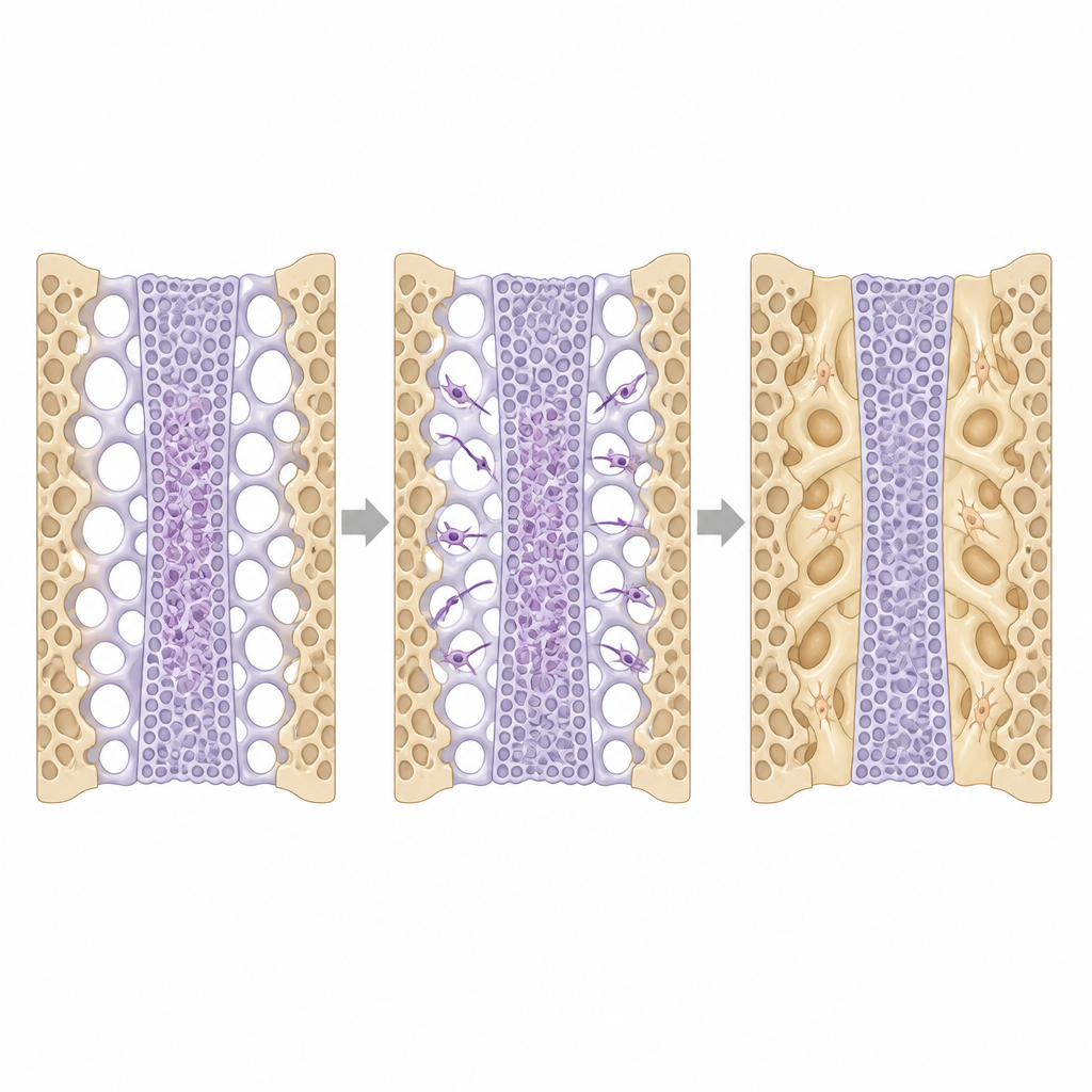
Guiding cell behavior and resisting unwanted bone
In cell and animal studies, the scaffold behaved as intended. When mixtures of naive stem cells and bone-forming cells were added, the more mature cells tended to migrate outward into the large-pore regions, where they laid down bone-like tissue. The naive cells stayed mostly in the small-pore center and showed markers of “stemness.” The central region also attracted fewer blood vessels and formed a less mature matrix, similar to a natural suture. Even when the researchers flooded the area with strong bone-forming signals in special mouse models, the central zone of the triphasic scaffold resisted filling in with solid bone, while the outer regions still healed into the surrounding skull.
Testing the idea in a disease model
To see if this could actually improve skull shape, the team turned to mice engineered to develop a common form of midline craniosynostosis. These animals show early fusion of a front skull seam and a characteristically short, wide face. The researchers developed a precise surgical procedure to remove the fused seam and insert the triphasic scaffold loaded with the animal’s own bone marrow stem cells. When this was done during a critical postnatal growth window, the implanted scaffold maintained an open, suture-like tissue and prevented the bones from re-fusing across the center. Detailed 3D measurements showed that treated mutant mice developed skull shapes much closer to normal, especially when treated earlier in the growth period.
From mouse skulls to future patient care
By combining clever scaffold design with the body’s own stem cells, this work shows that it may be possible to rebuild a functioning skull seam instead of repeatedly cutting bone. In mice, a temporary, biodegradable “bone–suture–bone” implant was enough to guide skull growth back toward a healthier pattern during a key developmental window. While much remains to be done before human use, the study offers a clear proof of concept that recreating a stem cell niche could one day reduce the need for large reconstructive surgeries in children with craniosynostosis.
Citation: Benton Swanson, W., Douglas, L., Woodbury, S.M. et al. A tissue engineering approach to regenerate the cranial suture skeletal stem cell niche with a multicompartment biomaterial scaffold. Bone Res 14, 58 (2026). https://doi.org/10.1038/s41413-026-00539-z
Keywords: craniosynostosis, cranial suture, tissue engineering, stem cell niche, biomaterial scaffold