Clear Sky Science · en
ER-phagy receptors: structural mechanisms in selective ER degradation and disease implications
Keeping the Cell’s Workshop in Balance
Every cell contains a sprawling inner workshop called the endoplasmic reticulum, or ER, where proteins are made, fats are processed and vital signals are managed. Like any busy factory, this workshop produces waste and damaged parts that must be cleared away. This review explains how cells use a special clean-up system, dubbed ER-phagy, to selectively remove worn-out pieces of the ER, and how failures in this system are linked to brain disorders, cancer, heart disease, metabolic illness and infections.
How Cells Recycle Their Inner Factory
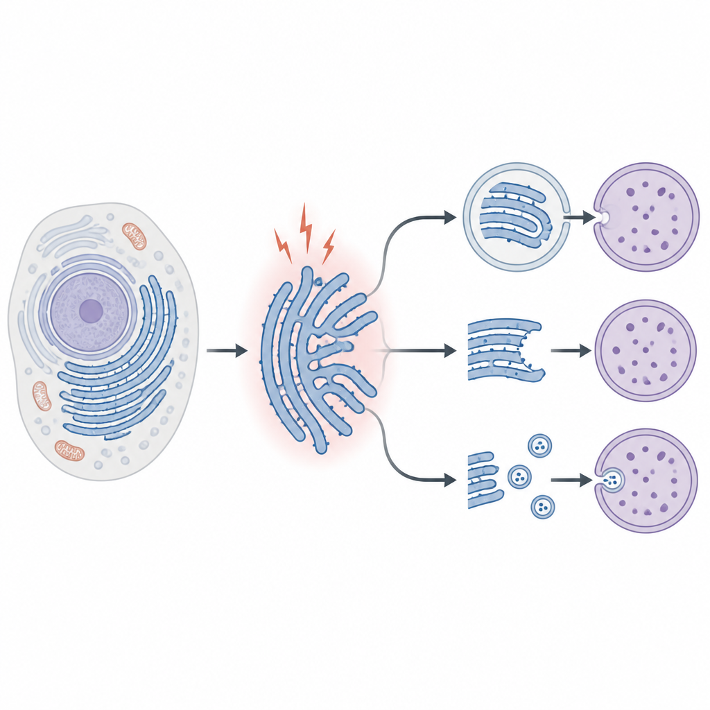
The ER is a maze of membranes that constantly adapts to the cell’s needs. When protein folding goes wrong or the cell faces stress, faulty proteins and damaged ER segments build up, threatening the whole operation. Cells respond with a broader self-eating program called autophagy, which recycles unwanted material. ER-phagy is the branch of this program that focuses specifically on the ER. It works through several routes: in macro-ER-phagy, bubbles with two membranes envelop chunks of ER and deliver them to cellular “stomachs” known as lysosomes; in micro-ER-phagy, lysosomes pinch off and swallow ER directly; and in a third route, ER-derived vesicles fuse with lysosomes without forming full autophagosomes. A newly described secretory pathway can even send ER fragments outside the cell instead of digesting them.
Special Gatekeepers on ER Membranes
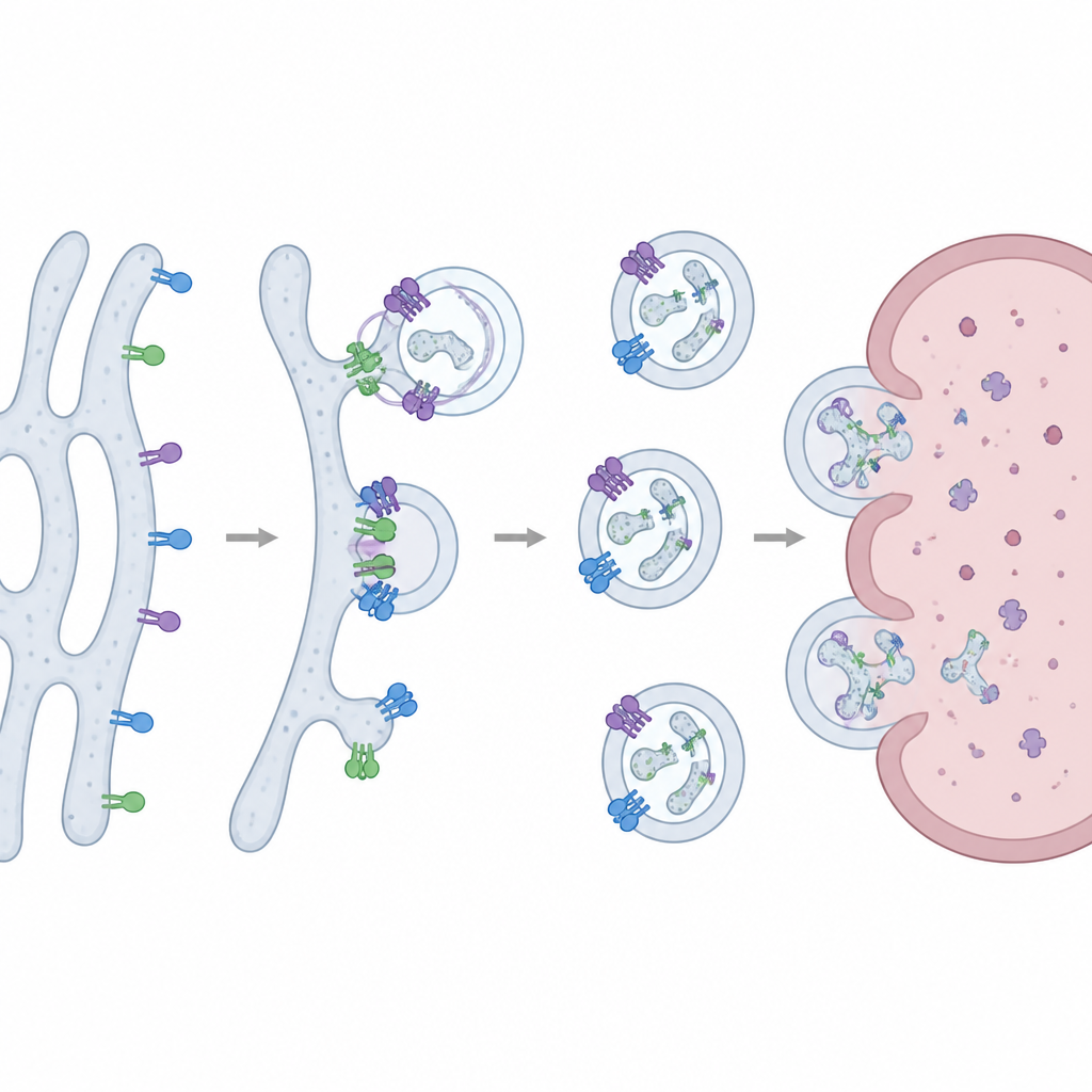
Central to ER-phagy are gatekeeper proteins called receptors that sit in or near the ER membrane and decide which parts should be removed. In yeast, two such receptors, Atg39 and Atg40, were first identified. In mammals, a larger cast has emerged, including FAM134 family members, RTN3L, TEX264, SEC62, CCPG1, ATL3, CALCOCO1 and others. Many have curved membrane–shaping segments that help pinch off ER fragments, along with flexible tails that latch onto autophagy proteins like LC3 and GABARAP. These tails act as tethers, linking marked ER regions to forming autophagy vesicles. Chemical tags such as phosphorylation and ubiquitination control when receptors cluster, how strongly they bind partners and whether they foster gentle housekeeping or aggressive breakdown.
Watching ER Cleanup in Action
Because ER-phagy is highly dynamic and occurs on tiny scales, scientists use several complementary tools to track it. Electron microscopy can directly reveal ER segments captured inside autophagosomes, offering striking visual proof but limited speed and throughput. Biochemical assays like Western blots measure changes in key ER markers and receptors as they are degraded, giving a bulk readout of activity. Fluorescent reporters that glow differently in neutral versus acidic compartments allow real-time imaging of ER pieces as they travel into lysosomes. New designer probes react to changes in viscosity, acidity or stress signals within the ER, providing sensitive ways to follow ER-phagy in living cells and even in complex disease models.
Links to Brain, Cancer, Metabolic and Heart Diseases
As researchers uncover these mechanisms, it has become clear that ER-phagy is deeply woven into human health. In the nervous system, receptors such as FAM134B, RTN3L and TEX264 help clear misfolded proteins, limit stress and support neuron development. Faulty or overactive ER-phagy receptors are tied to hereditary sensory neuropathy, Alzheimer’s disease, Parkinson’s disease, epilepsy and spinal cord disorders. In cancer, ER-phagy can either aid tumor cells by easing stress and supporting growth or push them toward death when overactivated. Certain receptors, like SEC62 and FAM134B, are overexpressed in some tumors and shape drug resistance, while others such as RTN3 and TEX264 may act as tumor suppressors. In metabolic and cardiovascular diseases, ER-phagy influences fat handling, hormone processing, diabetic organ damage and responses to heart injury. It also has surprising roles in infection, where it can either restrict viruses and bacteria or be hijacked by them.
From Cellular Housekeeping to Future Therapies
Taken together, the work reviewed in this article shows that ER-phagy is not a simple waste chute but a finely tuned quality-control network. Different receptors patrol distinct regions of the ER and can even form new tubular structures that divert proteins along unconventional secretory routes instead of toward destruction. When this network is balanced, cells keep their inner workshop healthy; when it is disturbed, chronic stress and disease can follow. Understanding which receptors matter most in each illness, and learning how to nudge their activity up or down, may open new ways to treat neurodegeneration, cancer, heart and metabolic diseases, and infections by restoring the cell’s own capacity to repair and reshape its vital ER factory.
Citation: Yang, Wj., Sheng, R. ER-phagy receptors: structural mechanisms in selective ER degradation and disease implications. Acta Pharmacol Sin 47, 1385–1400 (2026). https://doi.org/10.1038/s41401-025-01724-2
Keywords: autophagy, endoplasmic reticulum, cellular quality control, neurodegeneration, cancer biology