Clear Sky Science · en
sCMOS-based fNIRS system: validation via optical performance and cortical response
Seeing Brain Activity with Gentle Light
Imagine watching the brain at work without loud scanners or tight tubes, using only soft red light and a small camera. This study introduces a new way to do just that. The researchers tested a more compact and potentially cheaper brain imaging system that could one day help doctors and scientists study thinking, mood, and mental illness in more natural, everyday settings.
Why Soft Light is Used to Watch the Brain
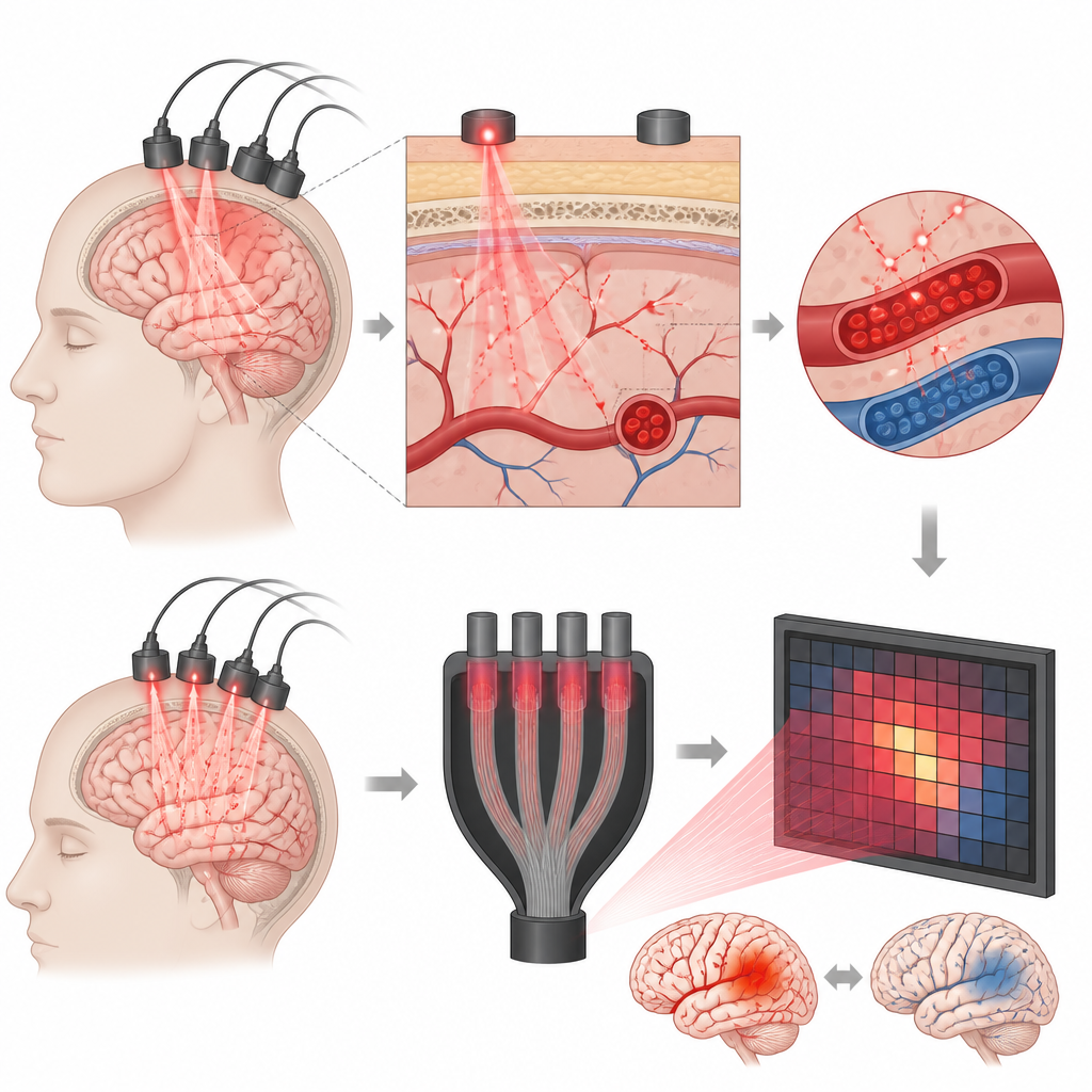
Functional near infrared spectroscopy, or fNIRS, shines near infrared light into the head and measures the faint light that comes back out. Because oxygen rich and oxygen poor blood absorb this light differently, tiny changes in blood flow linked to brain activity can be tracked over time. fNIRS is quiet, safe, and can be used while people sit, talk, or move, which makes it attractive for studying children and people with psychiatric or neurological conditions. However, current fNIRS machines often rely on many separate light detectors, making them bulky, expensive, and hard to scale up to cover the whole head.
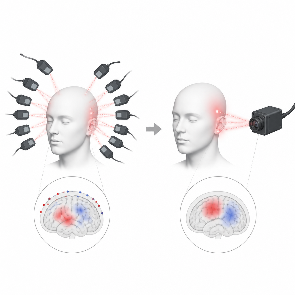
A New Camera Style Sensor for Brain Light
Traditional systems commonly use avalanche photodiodes, special light detectors that are very sensitive but require many individual units and support electronics. The authors instead built a system around a scientific CMOS camera sensor. Rather than having many separate detector units, they guide returning light through optical fibers onto different spots of a single two dimensional sensor chip. The setup includes near infrared laser diodes at two wavelengths, a grid of sources and detectors on a headpiece, optical filters to separate the two colors of light, and a control computer. By turning different light sources on and off in a timed pattern, the camera can tell which spot on the head each flash of light came from.
Testing Performance in Artificial “Tissue”
To find out whether this camera based system is as accurate as a standard fNIRS device, the team first tested it in carefully designed laboratory models that mimic how light travels through the head. One model mixed milky fluid and black ink to imitate scattering and absorption in layers like scalp, skull, and brain. By slowly adding tiny amounts of ink, they changed how strongly the mixture absorbed light, similar to how changing blood content would. The camera based system tracked these changes very precisely over a wide range, often more reliably than the conventional detector system, especially when signals were extremely weak or very strong. In a second model, they added real blood to a tissue like mixture and gradually removed oxygen from it. Both systems measured changes in blood oxygen in close agreement with a separate blood gas analyzer, showing that the new approach can follow realistic shifts in blood oxygen levels.
Watching Real Brains During a Thinking Task
The researchers next asked volunteers to perform a verbal fluency task, in which people quietly rest, then speak words from given categories, then rest again. This task is known to activate regions in the frontal part of the brain involved in language and executive function. Using a clever fiber splitter, the same returning light from the head was sent simultaneously to both the new camera based system and a commercial fNIRS machine. After cleaning the data and converting light changes into estimates of oxygenated and deoxygenated blood, the two systems produced very similar patterns over time in almost all channels, with strong statistical agreement. Occasional mismatches could be traced to practical issues like small shifts in a fiber position rather than to flaws in the sensor design.
What This Means for Future Brain Monitoring
In everyday terms, the study shows that a single smart camera chip can stand in for many separate light detectors without losing accuracy in measuring brain related blood flow signals. The camera based fNIRS system matched or outperformed a standard system across key measures of sensitivity, noise, stability over time, and response to realistic blood oxygen changes. Because it reduces size and complexity, this approach could help make brain monitoring tools more portable, less costly, and easier to adapt for people with different hair types and skin tones. While further engineering is needed to increase speed and shrink the hardware, this work suggests a clear path toward more accessible, camera powered brain imaging in both research and clinical care.
Citation: Zhou, J., Yan, B., Pu, Y. et al. sCMOS-based fNIRS system: validation via optical performance and cortical response. Transl Psychiatry 16, 260 (2026). https://doi.org/10.1038/s41398-026-03992-w
Keywords: functional near infrared spectroscopy, brain imaging, sCMOS sensor, cerebral blood oxygenation, psychiatric neuroimaging