Clear Sky Science · en
Gray and White matter microstructural alterations in major depressive disorder: a multi-center diffusion imaging study
Why Brain Wiring Matters in Depression
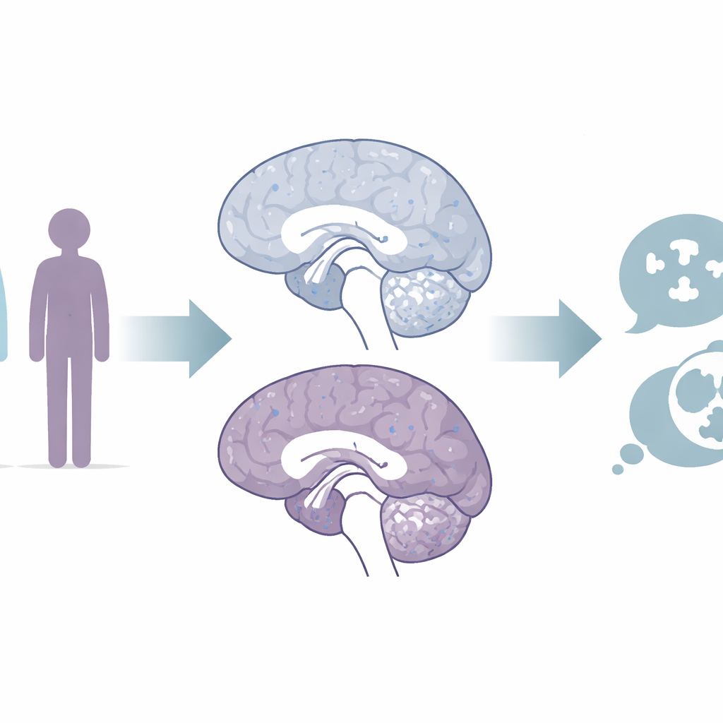
Major depression is often described in terms of feelings—persistent sadness, loss of interest, and exhaustion—but behind these experiences lies the physical machinery of the brain. This study set out to answer a simple but far-reaching question: does long‑term depression leave a trace in the brain’s fine wiring, not just in one or two "hot spots," but across gray and white matter throughout the brain? Using advanced MRI methods on a large group of patients from multiple hospitals, the researchers looked for subtle changes in brain tissue that might reflect inflammation, damage to connections, or other hidden stress on neural circuits.
Looking Inside the Brain’s Hidden Architecture
The team scanned the brains of 159 adults with major depressive disorder and 112 people without depression, using powerful 3‑tesla MRI machines at two centers in Japan. They focused on two kinds of brain tissue. Gray matter holds the cell bodies and branching processes that handle thinking and emotion. White matter contains the long, insulated fiber bundles that link distant regions into working networks. Traditional diffusion tensor imaging tracks how water moves through tissue to infer the health of these structures, but it struggles with the brain’s complexity. The researchers therefore combined it with a newer approach, neurite orientation dispersion and density imaging, which can tease apart different water compartments and provide a more detailed picture of how tightly packed and how orderly nerve fibers and branches are.
Signs of Extra Fluid and Fraying Connections
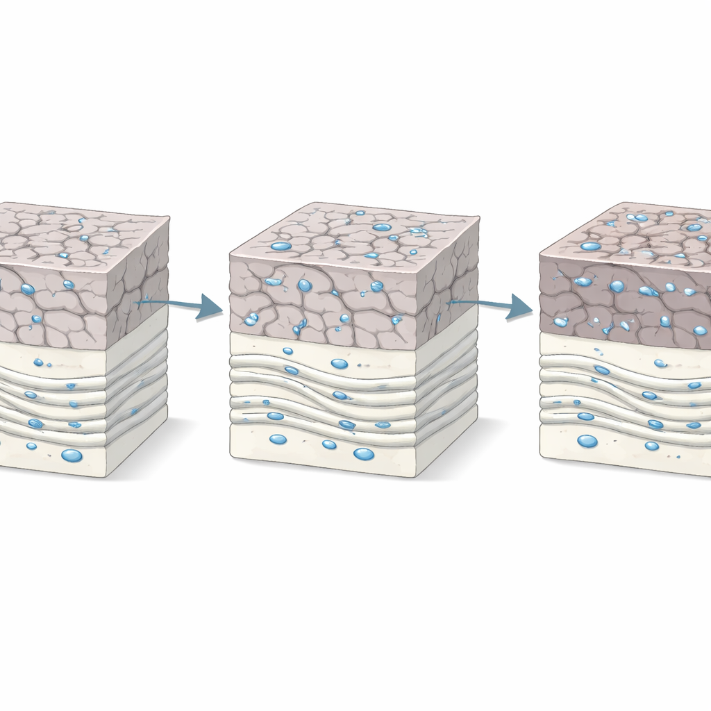
Across the brain’s gray matter, patients with depression showed a higher fraction of “free” water—water that is not tightly confined inside or between cells. This pattern was especially clear in regions deeply involved in mood and thinking, including the frontal and temporal lobes, the insula, hippocampus, and amygdala. In white matter, patients showed lower fractional anisotropy, a measure that typically falls when nerve fibers or their insulating myelin become less intact or less well aligned. They also showed higher orientation dispersion, suggesting that fiber directions were more disorganized overall. Additional diffusion measures pointed in the same direction, indicating that water could move more freely across the insulating layers of white matter, a pattern often linked to demyelination or inflammation.
Changes That Build Up Over Time
Interestingly, these tissue changes did not simply track how depressed people felt on the day of the scan. The imaging measures were not tied to scores on a standard depression rating scale. Instead, they related to how long a person had been living with the illness. The longer the history of depression, the lower the white matter integrity and the greater the disorganization of fiber directions. This suggests that the longer the brain is exposed to the biological stresses that accompany depression—such as elevated stress hormones or chronic inflammatory signals—the more its wiring may gradually fray. The gray matter increases in free water, meanwhile, lined up with regions previously shown to shrink in volume or function abnormally in depression, hinting that they may reflect inflammatory processes within key mood and memory hubs.
What This Means for Understanding Depression
Taken together, the findings paint depression not only as a disorder of mood, but as a condition in which the brain’s microstructure becomes subtly disorganized across many regions. Extra free water in gray matter and disrupted white matter pathways are consistent with ongoing neuroinflammation and damage to the insulating layers around nerve fibers. Because these changes are widespread and related to how long a person has been ill, they may help explain why some symptoms—such as difficulties with concentration, motivation, and memory—can become more stubborn over time. While the scans cannot yet be used as a simple diagnostic test, they offer important clues that future work can connect to cellular and molecular changes, potentially guiding more targeted treatments aimed at protecting or repairing the brain’s wiring in depression.
Citation: Takahashi, K., Suwa, T., Yoshihara, Y. et al. Gray and White matter microstructural alterations in major depressive disorder: a multi-center diffusion imaging study. Transl Psychiatry 16, 163 (2026). https://doi.org/10.1038/s41398-026-03916-8
Keywords: major depressive disorder, brain microstructure, diffusion MRI, white matter, neuroinflammation