Clear Sky Science · en
Patient-derived brain organoids reveal divergent neuronal activity across subpopulations of autism spectrum disorder
Peeking Into the Developing Human Brain
Autism is famously complex: two people with the same diagnosis can think, feel, and behave in very different ways. This study uses tiny lab-grown “mini-brains,” called brain organoids, made from cells donated by autistic and non-autistic individuals, to see how their neurons fire and talk to each other. By comparing electrical activity across different genetic forms of autism and typical controls, the researchers hope to uncover hidden patterns in brain wiring that could someday guide diagnosis and treatment.
From Urine Sample to Mini-Brain
The journey in this study starts with something as ordinary as a urine sample. Cells from urine were reprogrammed into induced pluripotent stem cells—cells that can become many types of tissue. These were then guided to form three-dimensional brain organoids, tiny spheres that mimic early human brain development. The organoids contained a mix of nerve cells and support cells resembling those found in the developing cortex, including both excitatory and inhibitory neurons. The team confirmed this by profiling gene activity cell by cell and by staining for hallmark proteins that mark immature and more mature neurons. 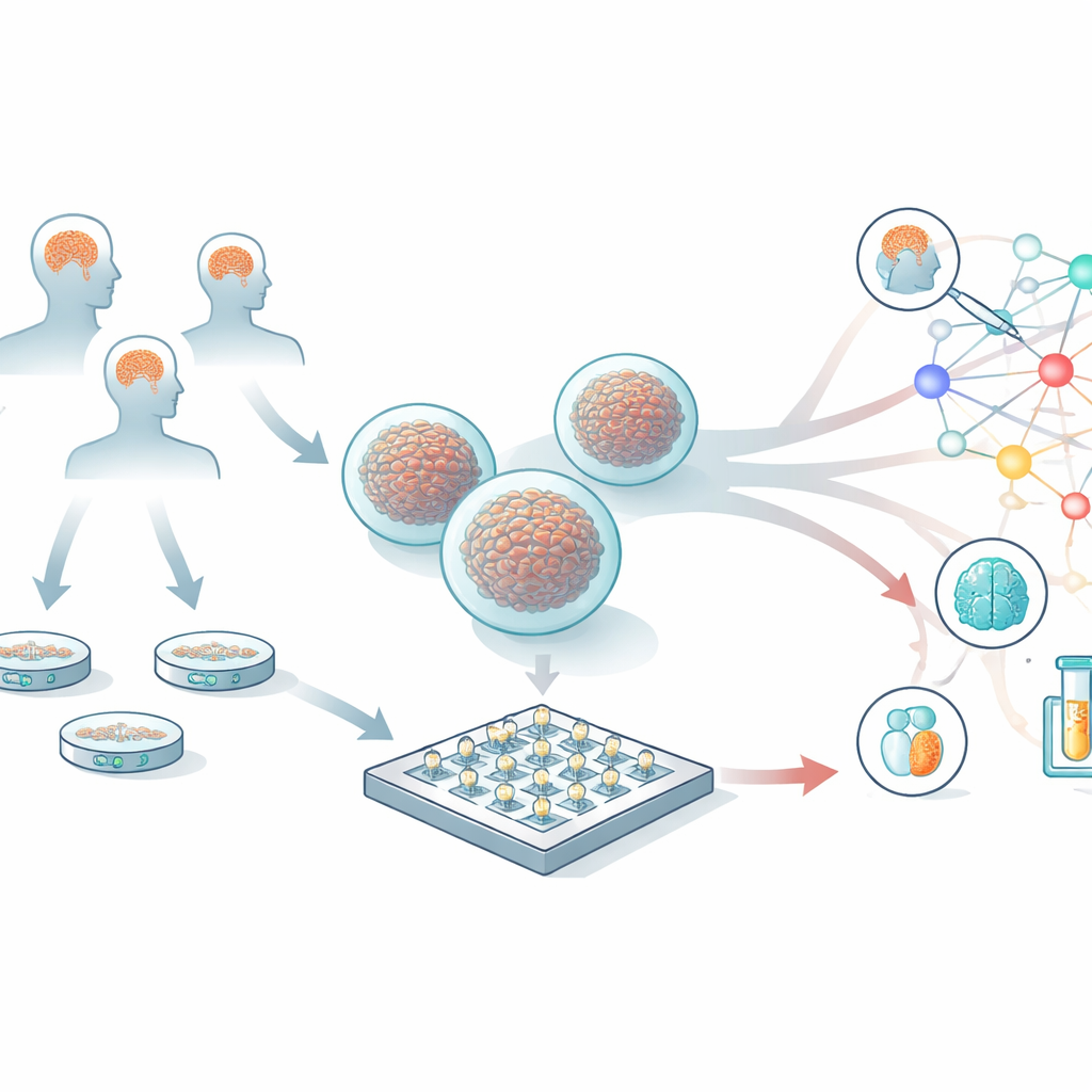
Different Electrical Voices in Autism
The researchers studied organoids from eleven people with autism—ten with known single-gene syndromes and one with idiopathic (non-syndromic) autism—alongside organoids from four neurotypical individuals. When they listened to the baseline, or “resting,” activity, clear differences emerged. Organoids from the idiopathic autism donor were generally quiet, with fewer electrical spikes and bursts than controls. In contrast, several genetic autism groups, including those with changes in STXBP1, PPP2R5D, and GRIN2B, showed heightened firing rates, like a network running “too hot.” Even within the same genetic syndrome, different individuals could show distinct firing patterns, echoing the clinical reality that the same gene change does not always lead to the same symptoms.
How Mini-Brains Respond to a Jolt
Real brains constantly adjust their responses to incoming signals, a property known as plasticity. To mimic this, the team gave the organoids brief bursts of electrical stimulation and then measured how their firing patterns changed. In most organoids, these rapid pulses led to a short-term dampening of activity, a kind of built-in braking system. But the balance between strengthening and weakening responses varied strikingly across autism subtypes. Some genetic forms, such as STXBP1, SHANK3, and one SCN2A line, showed exaggerated depression of activity and reduced strengthening, suggesting their networks were unusually prone to “shutting down” after a burst. GRIN2B organoids, on the other hand, showed a more balanced or even slightly enhanced strengthening response, hinting at a distinct way their synapses adapt to input.
Network Wiring Under Stress
The study also looked beyond individual spikes to the larger communication web—how many electrodes were talking to each other and how strongly. In control organoids, the functional network was fairly stable, with a modest, consistent reduction in connectivity after stimulation. Autism-derived organoids told a more varied story: some showed a sharp collapse in network size, others an erratic or blunted response, and still others started off with unusually dense connectivity that broke down after stimulation. These differences suggest that the way neural circuits organize and reorganize themselves in response to challenges is altered in autism, and in ways that depend on the specific gene involved.
Pulling Many Signals Into One Picture
To make sense of the 18 different electrical features they measured—from firing rate and burst frequency to network density—the researchers used a statistical technique that compresses complex data into a three-dimensional map. In this map, organoids from the same person clustered closely together, showing that the method captures stable, individual “signatures.” Control donors formed a tight group, while autism organoids spread out over a much broader space. Each genetic subtype tended to occupy its own region, but with overlap and internal diversity. This pattern reinforces the idea that autism is both many conditions and one: different gene changes can push brain networks away from typical function in distinct yet partly converging ways. 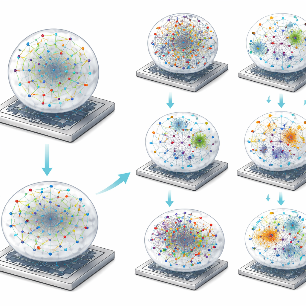
What This Means for Understanding Autism
In plain terms, this work shows that tiny lab-grown brain models can capture real and meaningful differences in how neurons from autistic and non-autistic people fire, adapt, and wire together. Rather than a single “autism brain pattern,” the study reveals a landscape of electrical behaviors: some networks are overactive, some underactive, some fragile after stimulation, and some oddly rigid. Yet these diverse routes often lead to a common outcome—disrupted communication in brain circuits. By linking specific genetic changes to particular electrical fingerprints, patient-derived brain organoids could become a powerful tool for earlier diagnosis, for sorting patients into biologically informed subgroups, and for testing which experimental drugs restore more typical network activity.
Citation: Perets, N., Kerem, L., Waiskopf, N. et al. Patient-derived brain organoids reveal divergent neuronal activity across subpopulations of autism spectrum disorder. Transl Psychiatry 16, 164 (2026). https://doi.org/10.1038/s41398-026-03890-1
Keywords: brain organoids, autism spectrum disorder, neuronal networks, synaptic plasticity, personalized neuroscience