Clear Sky Science · en
Synaptic dysfunction and adaptation after NMDA receptor ablation in the mouse medial prefrontal cortex
Why Brain Wiring in Teen Years Matters
Adolescence is a period when the brain’s wiring is heavily remodeled, and this remodeling is thought to be linked to the appearance of mental illnesses such as schizophrenia. The study summarized here asks a simple but powerful question: what happens to the fine connections between brain cells in the thinking centers of the brain if a key type of communication channel is gradually shut down during the teenage period? Understanding how the brain first falters and then adapts could reveal why some people develop lasting psychiatric symptoms while others recover.
A Focus on a Critical Brain Hub
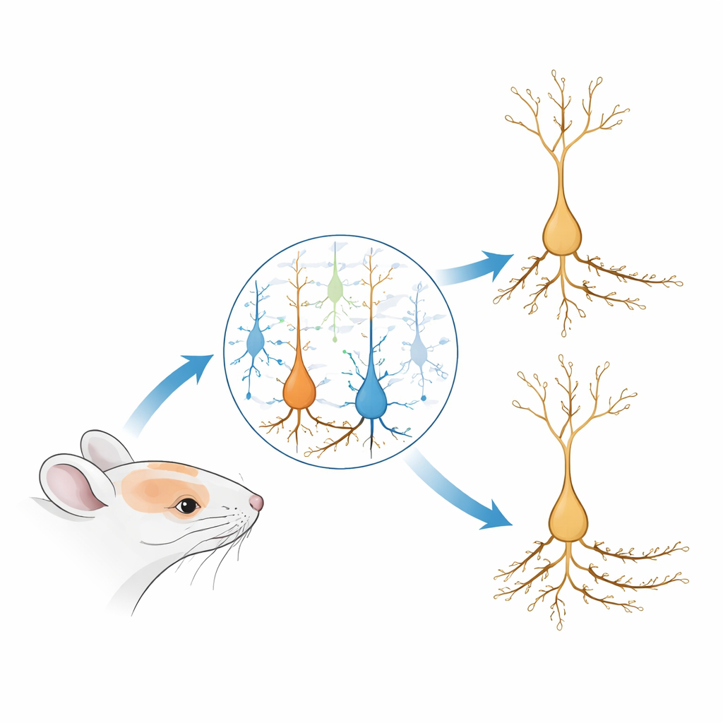
The researchers zoomed in on the medial prefrontal cortex of mice, a region important for decision-making, working memory, and flexible thinking—abilities that are often disrupted in schizophrenia. They focused on NMDA receptors, molecular gatekeepers on neurons that help tune the strength of connections and support coordinated firing across brain networks. Drugs or autoimmune attacks that block these receptors can temporarily produce schizophrenia-like symptoms, and people with the illness often show altered NMDA receptor function and fewer tiny contact points, called dendritic spines, on prefrontal neurons. However, it has been unclear how a slow loss of these receptors specifically during adolescence reshapes the wiring and activity of local circuits.
Editing a Single Gene in the Adolescent Brain
To probe this, the team used a CRISPR-based gene-editing strategy in adolescent mice. They delivered a virus into the medial prefrontal cortex that turned on a DNA-cutting enzyme together with a guide sequence targeting Grin1, the gene needed to build the essential subunit of NMDA receptors. This allowed them to progressively eliminate NMDA receptors from many neurons in that region while leaving the rest of the brain intact. Using electrical recordings in brain slices, they confirmed that signals carried by NMDA receptors were strongly reduced, reaching levels similar to what is seen when receptors are fully blocked by drugs. At the same time, they filled individual neurons with a tracer and used high-resolution confocal microscopy to reconstruct their branching trees and count spines along different parts of the cell.
A Two-Phase Change in Tiny Connections
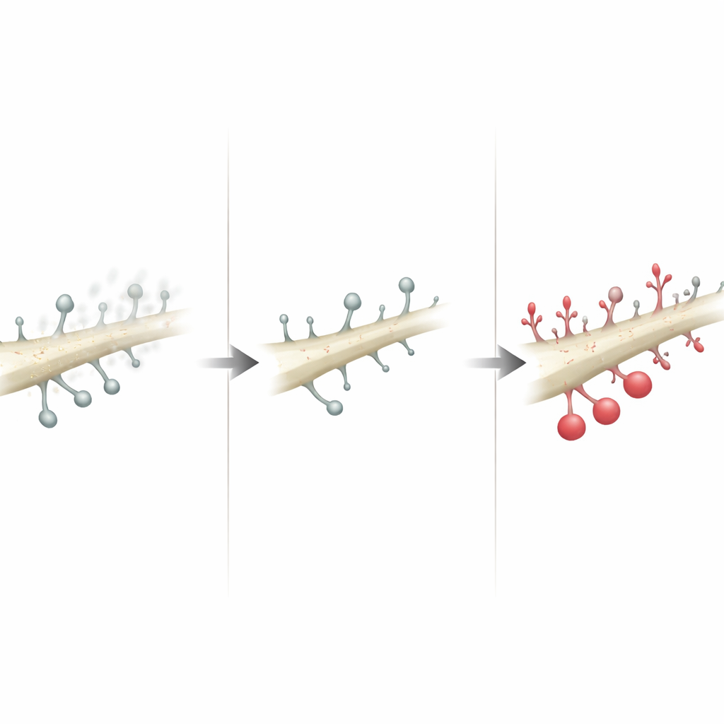
The team discovered that the small branches emerging from the lower part of the neuron—the basilar dendrites—underwent a striking two-phase change. A couple of weeks after gene editing, these branches had fewer spines, mainly due to a loss of the smallest, most fragile protrusions and thin filaments thought to represent weak or nascent contacts. By six weeks, however, this pattern flipped: basilar branches actually carried more spines than in control animals, especially in the less mature categories. In contrast, the long upper branch of the cell—the apical dendrite, which receives more distant inputs—showed no consistent changes. This suggests that local connections among nearby neurons in the prefrontal cortex are especially sensitive to NMDA receptor loss, but they can also mount a robust comeback.
How Electrical Signals Adjust Over Time
These structural shifts were mirrored by changes in spontaneous electrical events. Early on, the strength of individual excitatory events did not change, and their frequency remained similar to controls, despite the initial drop in spine numbers. By six weeks, though, excitatory events occurred more often, consistent with the overshoot in spine density, while their average size stayed the same. The researchers ruled out several simple explanations: the probability that sending neurons released their chemical messenger did not change, and the make-up of the main fast receptors for that messenger appeared stable. At the same late stage, they also detected stronger inhibitory events arriving onto pyramidal neurons, suggesting that inhibitory cells had adjusted their output upward. Together, these results point to a network that rebalances itself after the loss of NMDA signaling by adding more excitatory contacts while also boosting inhibition.
When Cell Type and Risk Factors Matter
To test whether these adaptations were driven solely by the main excitatory neurons, the team repeated the gene-editing experiment using a promoter that largely restricts expression to those cells. In this more selective manipulation, NMDA signals in the targeted neurons were reduced, but there were no clear changes in spine density or excitatory events. This implies that broader loss of NMDA receptors across multiple cell types—or especially within inhibitory cells—may be required to trigger the cascading reorganization seen with the pan-neuronal manipulation. The authors connect these findings to human studies showing altered inhibitory signaling and reduced markers of certain interneurons in schizophrenia, and they suggest that a complex interplay of excitatory and inhibitory cells may underlie both vulnerability and compensation in the disorder.
What This Means for Mental Health
From a lay perspective, the main message is that when a key signaling pathway in the prefrontal cortex is weakened during the teen years, the local wiring first thins out and then regrows in a altered pattern, with both excitatory and inhibitory signals adapting to reach a new balance. This rebound indicates that the adolescent brain has a strong ability to compensate for certain molecular disruptions, which may help explain why some individuals with risk-related changes never develop chronic symptoms. At the same time, the study suggests that if other genetic or environmental factors also limit the capacity to regrow and stabilize these tiny connections, the system may fail to recover, contributing to lasting cognitive problems. Understanding these adaptive and maladaptive responses opens the door to therapies that support healthy rewiring and help restore balanced signaling in psychiatric disorders.
Citation: Dick, R.M., Cunitz, L.B., Torres Pérez, A. et al. Synaptic dysfunction and adaptation after NMDA receptor ablation in the mouse medial prefrontal cortex. Neuropsychopharmacol. 51, 1100–1109 (2026). https://doi.org/10.1038/s41386-026-02381-7
Keywords: NMDA receptor hypofunction, medial prefrontal cortex, dendritic spine plasticity, schizophrenia risk, adolescent brain development