Clear Sky Science · en
Effects of electroconvulsive shock on the function, circuitry, and transcriptome of dentate gyrus granule neurons
Shocking the brain to lift mood
Electroconvulsive shock, the laboratory version of electroconvulsive therapy, is one of the most effective treatments for severe, treatment‑resistant depression, yet how it helps the brain recover has remained mysterious. This study in mice looks deep inside a memory‑related brain region called the hippocampus to find out how repeated shocks change brain cells, their wiring, and their gene activity in ways that may ease anxiety and depression‑like behavior.
From stressed mice to eased behavior
The researchers first created a long‑lasting stress state in mice by adding the stress hormone corticosterone to their drinking water, a common model for depression‑like behavior. After several weeks, mice received either a series of electroconvulsive shocks or sham treatment, then were tested in tasks that reflect anxiety and despair‑like states. Stressed mice that received shocks approached food faster in a new, mildly threatening arena and struggled longer in a forced swim test, both signs of reduced anxiety‑ and depression‑like behavior, while overall movement stayed normal. These changes mirror the clinical picture in which repeated shock treatments can help patients who do not respond to standard antidepressant drugs. 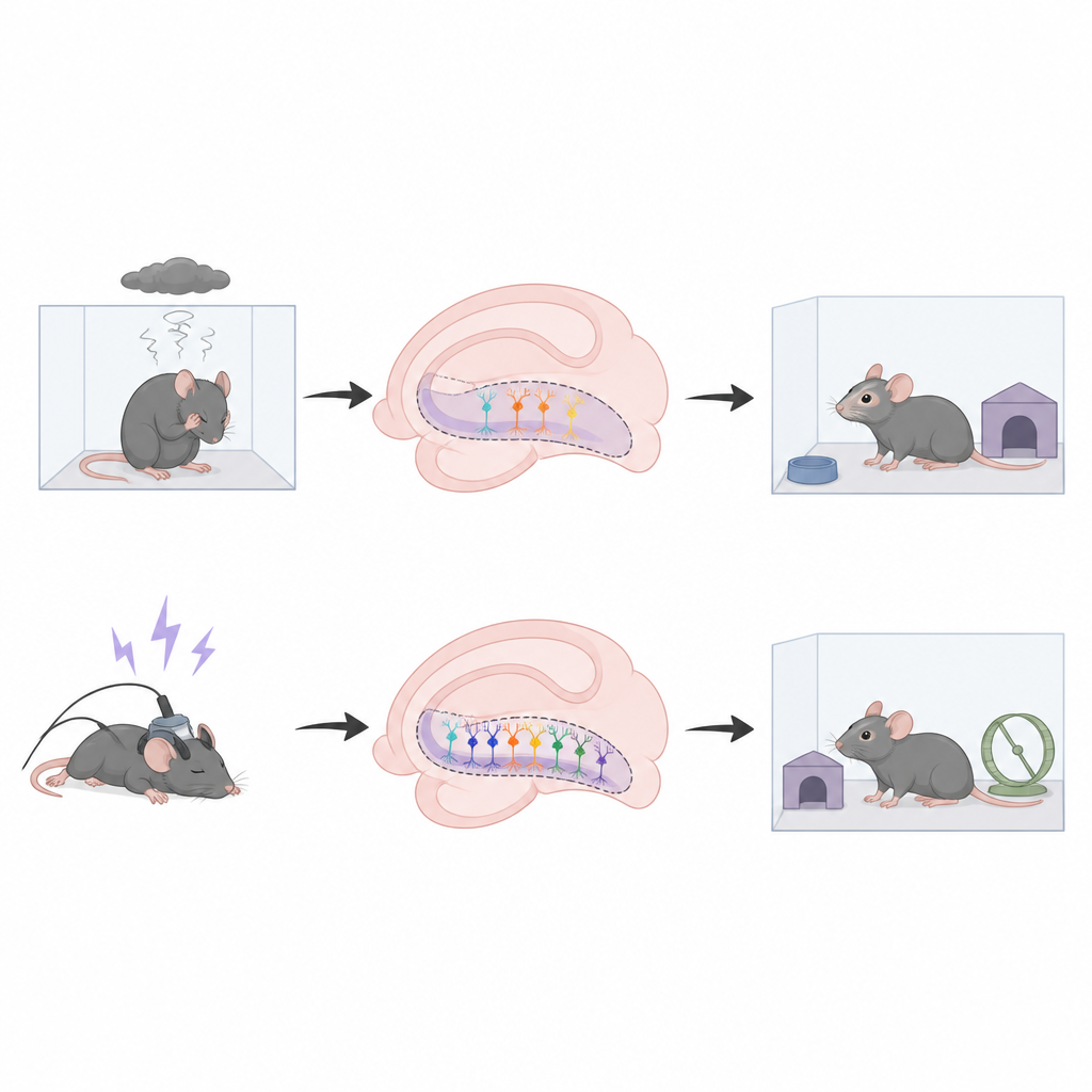
New brain cells as hidden helpers
Attention then turned to tiny newborn neurons in a part of the hippocampus called the dentate gyrus. Using a protein marker that tags immature cells, the team showed that repeated shocks, but not a single shock, boosted the number of these young neurons along the full length of this brain area. To test whether these cells are actually needed for the helpful behavioral effects, the scientists used targeted X‑ray treatment to wipe out new neuron growth in the dentate gyrus before giving shocks. In mice lacking new neurons, shocks no longer reduced anxiety‑ or depression‑like behavior, indicating that adult‑born cells are a critical link between treatment and mood improvement.
Quieting overactive circuits
New neurons in this region are known to be surprisingly active yet help maintain overall quiet, sparse firing of the surrounding network. The authors found that after a course of shocks, mature dentate gyrus cells showed fewer signs of recent activity at rest, suggesting a calmer circuit. With fine‑scale electrical recordings in brain slices, they stimulated young neurons with light and measured how strongly they could silence mature neighbors. After shock treatment, activation of immature cells caused a larger hyperpolarizing signal in mature cells, an effect that depended on a specific type of glutamate receptor. Blocking this receptor removed the extra inhibition, supporting the idea that shocks strengthen a pathway in which young cells directly quiet older ones and help prevent runaway activity linked to stress.
Gene activity shifts toward a youthful state
Finally, the team examined gene readouts from thousands of individual hippocampal neurons using single‑nucleus RNA sequencing. They compared stressed mice to stressed mice treated either with shocks or with the antidepressant fluoxetine. Both treatments increased the share of granule cells showing an “immature‑like” gene pattern and boosted genes tied to growth, connectivity, and plasticity, while dialing down markers of fully mature cells. However, the overall gene‑expression fingerprints of shock and drug treatment were not the same: fluoxetine mainly turned many genes up, whereas shocks tended to turn many genes down, and each treatment affected distinct gene sets in multiple hippocampal cell types. 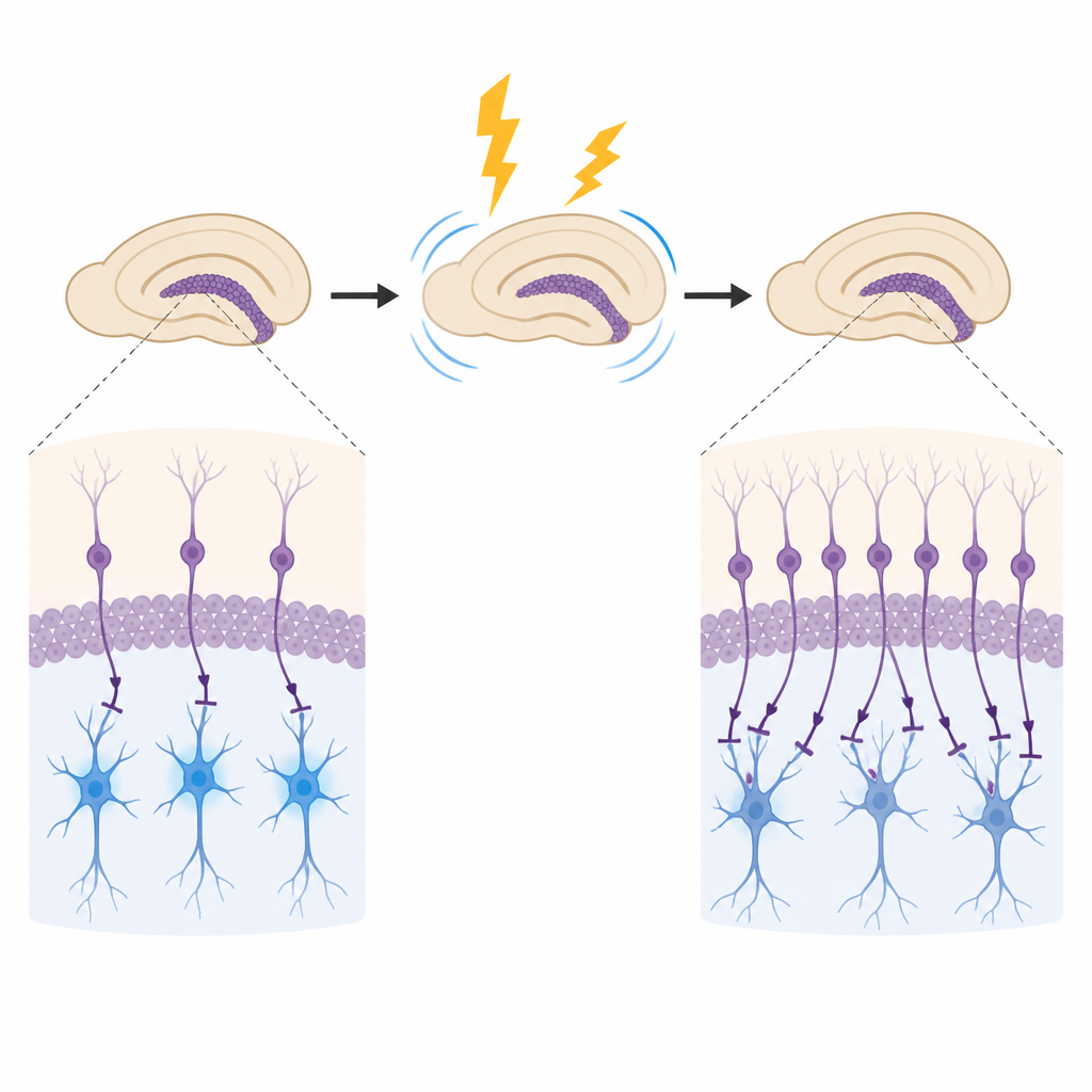
What this means for depression treatment
Taken together, the results suggest that the mood‑lifting effects of electroconvulsive shocks rely on a small but powerful pool of young hippocampal neurons. Repeated shocks increase the number of these cells, strengthen their ability to rein in older neighbors, and tilt gene programs in dentate gyrus neurons toward a more flexible, youthful state. Although both shocks and antidepressant drugs enhance plasticity, they do so in different molecular ways, which may help explain why shock treatment can work when medications fail and why it carries its own side effects. Understanding these cell‑level changes may guide future therapies that capture the benefits of electroconvulsive treatment while reducing its risks.
Citation: Santiago, A.N., Saval, J.C., Nguyen, P. et al. Effects of electroconvulsive shock on the function, circuitry, and transcriptome of dentate gyrus granule neurons. Neuropsychopharmacol. 51, 1258–1266 (2026). https://doi.org/10.1038/s41386-026-02345-x
Keywords: electroconvulsive therapy, hippocampal neurogenesis, dentate gyrus, stress resilience, antidepressant mechanisms