Clear Sky Science · en
Multiple sources of β2*-nicotinic acetylcholine receptor binding are differentially affected during tobacco smoking abstinence as revealed by Independent Component Analysis of [18F]Flubatine PET images
Why this research matters for people who smoke
Tobacco smoking remains one of the leading causes of preventable death, yet the brain changes that keep people hooked and make quitting so hard are still being uncovered. This study peeks inside the living human brain to see how different groups of nicotine sensitive receptors respond when people stop smoking. Understanding these hidden shifts may guide future treatments that make it easier to quit and stay smoke free.
Looking at nicotine’s footprints in the brain
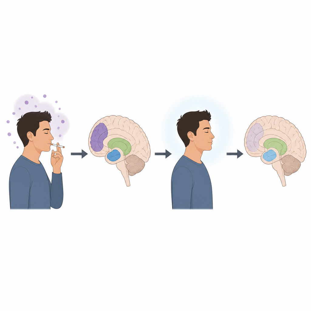
Nicotine exerts its effects by binding to nicotinic acetylcholine receptors, a family of proteins that act like tiny switches on brain cells. Many of these switches contain a building block called the beta2 subunit, but they can be combined with several other pieces to form slightly different versions with distinct jobs in behavior, mood, and thinking. Standard brain scans can see the overall presence of these receptors but struggle to tell one version from another. The researchers used a highly sensitive brain imaging tracer called [18F]Flubatine together with a data driven method called independent component analysis to tease apart separate patterns of binding that likely reflect different receptor combinations.
Separating hidden patterns of receptor binding
The team analyzed brain scans from people who had never smoked and from people who had recently stopped smoking cigarettes. Their method reliably revealed three distinct patterns of tracer binding. Two of them were centered in deep relay hubs of the brain called the thalamus and nearby midbrain regions, while the third pattern was strongest in the cerebellum, an area important for coordination and increasingly recognized in addiction research. When volunteers in a separate group took in nicotine from cigarettes or electronic cigarettes during scanning, the signal from all three patterns dropped, showing that each pattern reflected real nicotine sensitive receptors rather than background noise.
Pinpointing a receptor type rarely seen in living people
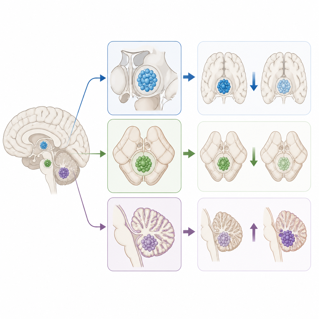
To figure out which specific receptor types these patterns might represent, the scientists turned to post mortem human brain tissue. They exposed thin slices of thalamus and cerebellum to the same tracer together with selective blocking compounds that each target different receptor subtypes. In the thalamus, blocking agents that prefer the common alpha4beta2 subtype displaced most of the tracer, supporting the idea that the two thalamic patterns arise largely from this subtype. In the cerebellum, however, a blocker with strong action at receptors containing the alpha3 subunit displaced the most tracer, while a blocker that favors alpha6 containing receptors had much weaker effects. This suggests that the cerebellar pattern largely reflects alpha3beta2 receptors, a subtype not previously isolated in living human brains.
How quitting smoking changes different receptor pools
When the researchers compared people who never smoked with people who had stopped smoking for about a week, clear differences emerged. The two thalamic patterns showed lower levels in abstinent smokers, and heavier daily smoking tended to be linked with even lower values. In contrast, the cerebellar pattern associated with alpha3beta2 receptors tended to be higher in people who had been abstinent and showed early signs of rising with both cigarettes per day and severity of nicotine dependence. At the same time, a broad measure of overall receptor availability across much of the brain was higher in abstinent smokers, echoing earlier work showing that many nicotine sensitive receptors are upregulated after chronic use.
What this means for helping people quit
Taken together, these findings suggest that different groups of nicotine sensitive receptors in the human brain do not all respond to smoking and abstinence in the same way. Some pools, especially in the thalamus, appear reduced in people who smoke, while those in the cerebellum that likely include alpha3beta2 receptors appear increased and linked to how much a person smokes and how dependent they are. By showing that it is possible to pick out the signal of this specific receptor subtype in living people, the study opens a path to test new medicines aimed at these targets. In the future, such advances could help design treatments that ease withdrawal symptoms, support thinking and mood, and ultimately improve the chances of quitting successfully.
Citation: Raval, N.R., Calakos, K.C., Zheng, MQ. et al. Multiple sources of β2*-nicotinic acetylcholine receptor binding are differentially affected during tobacco smoking abstinence as revealed by Independent Component Analysis of [18F]Flubatine PET images. Neuropsychopharmacol. 51, 1207–1216 (2026). https://doi.org/10.1038/s41386-025-02311-z
Keywords: nicotine receptors, smoking abstinence, brain imaging, PET scans, tobacco addiction