Clear Sky Science · en
Systems genetic dissection of brain gene expression reveals excitotoxic mechanisms of Alzheimer’s disease
Why this study matters for brain health
Alzheimer disease is often described as a buildup of harmful proteins in the brain, but how those deposits actually destroy nerve cells has remained unclear. This study connects the dots between those early changes and later memory loss by using fruit flies as tiny stand-ins for the human brain. By doing so, the researchers uncover a key role for runaway brain activity, where overstimulated nerve cells slowly injure themselves, and they pinpoint groups of genes that either worsen or soften this damage.
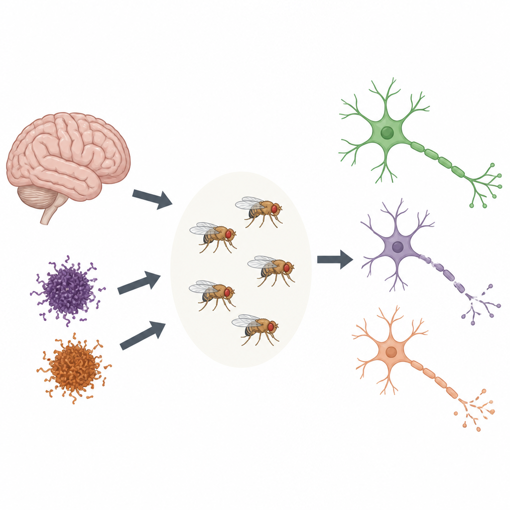
Turning flies into stand-ins for the aging brain
Instead of relying only on brain tissue donated after death, which captures just the final stage of disease, the team created fruit fly strains that produce the same toxic proteins seen in Alzheimer disease. Some flies made amyloid beta, which forms sticky plaques, while others made tau, the protein found in tangles inside nerve cells. The scientists tracked these flies over their life span, measuring both movement problems and changes in gene activity in their brains. Because flies age quickly and are easy to manipulate genetically, this approach let them watch how gene activity shifts from early life through later decline.
Finding gene networks that shape decline
The researchers compared the fly data with large catalogs of gene activity from thousands of human brains, where genes that switch on and off together form networks. They showed that most of the human Alzheimer-related networks have matching versions in flies, and that these shared networks respond to amyloid, tau, and normal aging. One set of networks involved the brain’s immune response, while another centered on communication between nerve cells at synapses. This cross-species overlap suggests that many of the same molecular systems are disturbed in both flies and people as the disease advances.
Testing which genes actually drive damage
To move from correlation to cause, the team systematically altered 344 high-priority genes that sit in key positions inside those human networks, using genetic tools in flies. They then asked whether turning each gene up or down made amyloid- or tau-driven nerve damage better or worse, as judged by the flies’ ability to climb and by visible holes in their brain tissue. This large-scale test revealed 141 “modifier” genes: some changes amplified the damage, while others clearly protected nerve cells. An immune-related network tended to contain genes whose increased activity sped up degeneration, hinting that a persistent inflammatory state in neurons may be harmful rather than helpful.
Runaway activity and the stressed synapse
One synapse-centered network, called PHGbrown in the human data, behaved in a more complex way. Many of its genes help nerve cells send and receive glutamate signals, the main “go” chemical in the brain. In people with Alzheimer disease this network is dialed down overall, but in early-stage models and certain cell types it is initially turned up. Using special calcium-sensitive reporters, the team showed that amyloid in flies leads to hyperactive neurons with excessive calcium entry, a hallmark of excitotoxic stress. By either reducing glutamate packaging into synaptic vesicles or dialing down selected PHGbrown genes, including the gene for a major glutamate receptor subunit, they could curb this overactivity and limit structural damage in the fly brain.
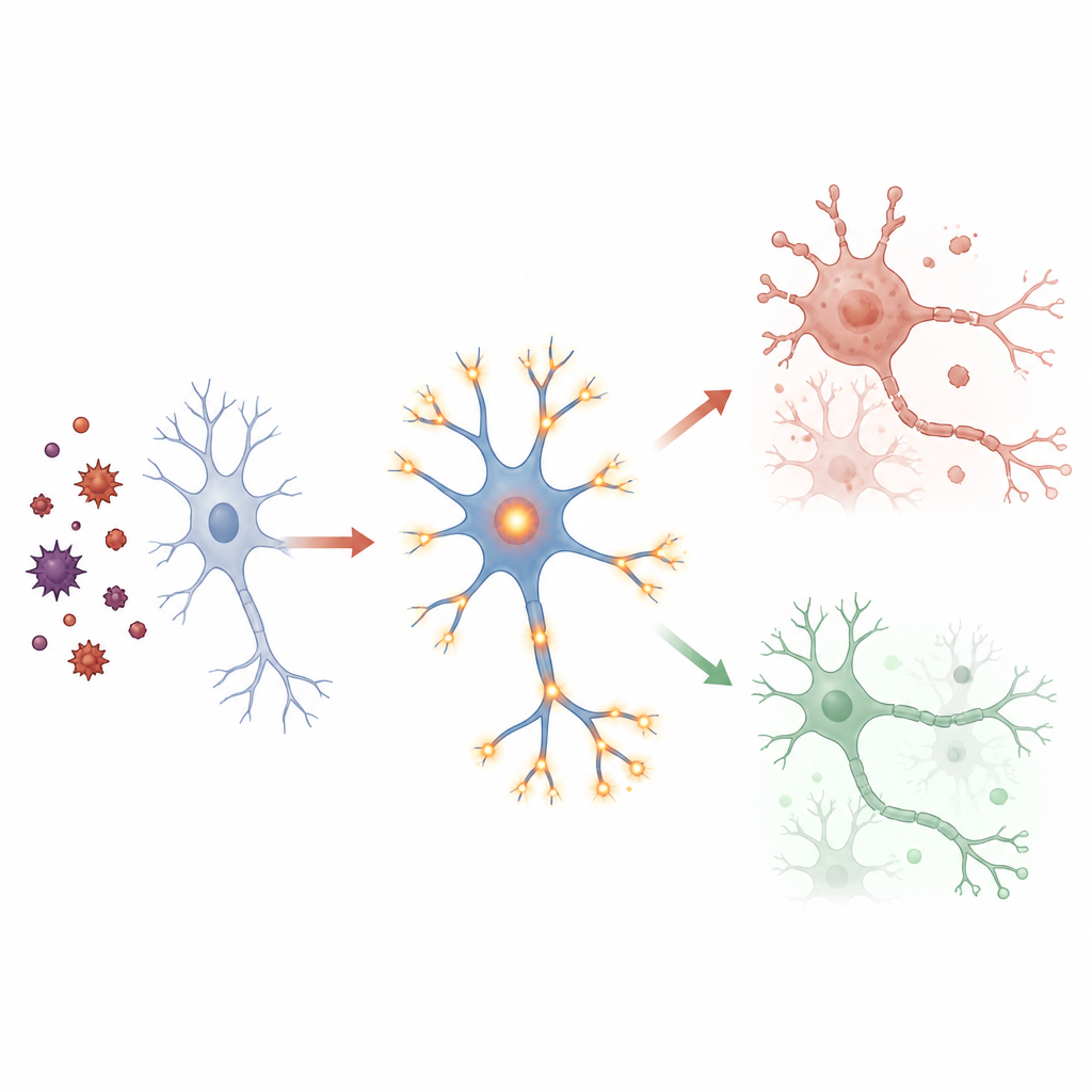
A two-phase story of stress and adaptation
Putting these pieces together, the authors propose a two-phase model. Early in the course of Alzheimer disease, amyloid appears to push nerve cells into an overexcited state, and the synaptic gene network ramps up, which ironically increases stress and injury. As damage accumulates and tau pathology grows, the same network is later dialed down, which may partly protect surviving neurons by dampening activity, even as some synapses and cells are lost. Their work links toxic proteins, altered gene activity, excessive glutamate signaling, and neuron death into a single causal chain, offering new ideas for therapies that aim not at a single protein, but at stabilizing the broader gene networks that keep brain activity in a healthy range.
Citation: Zhao, P., El Fadel, O., Le, A. et al. Systems genetic dissection of brain gene expression reveals excitotoxic mechanisms of Alzheimer’s disease. Mol Psychiatry 31, 3462–3481 (2026). https://doi.org/10.1038/s41380-026-03479-6
Keywords: Alzheimer disease, gene networks, excitotoxicity, synaptic function, fruit fly models