Clear Sky Science · en
Cerebellar microglia-derived IL-17A mitigates autism-related behavioral and synaptic deficits
Why brain immune cells matter for social behavior
Autism spectrum disorder affects millions of people worldwide, yet current treatments have limited impact on core symptoms such as social difficulties and repetitive behaviors. This study explores an unexpected ally inside the brain: tiny immune cells in the cerebellum that release a signaling molecule called IL-17A. By studying mice with a genetic form of autism-like traits, the researchers show how this molecule can actually help stabilize brain circuits involved in social behavior, hinting at new ways to relieve symptoms without broadly suppressing or overstimulating the immune system.
A small brain region with a big social role
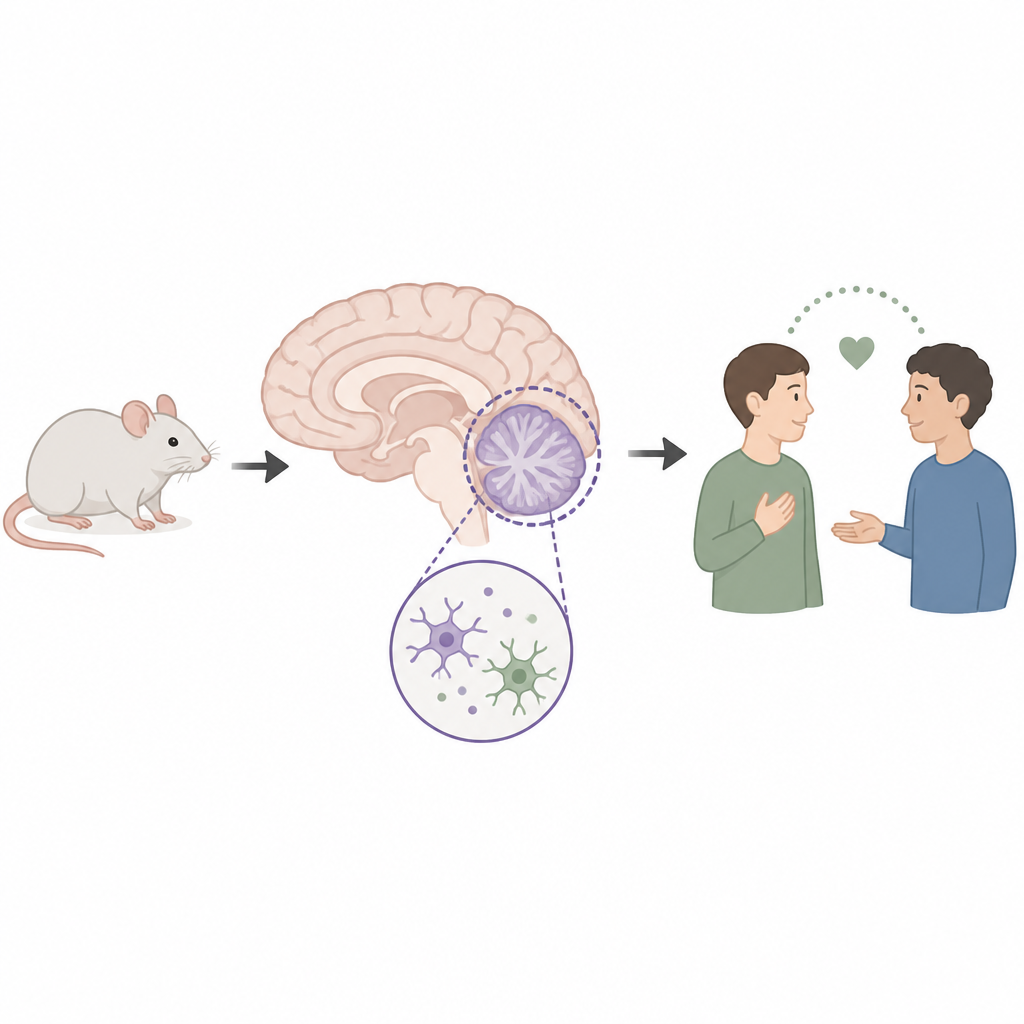
The cerebellum is usually associated with balance and movement, but growing evidence links one of its subregions, called Crus I, to social thinking and emotion. In mice lacking the Fmr1 gene, a widely used model of Fragile X syndrome and autism, the team first confirmed classic autism-like behaviors: poor interest in other mice and an increase in repetitive digging, captured by a marble-burying test. They then recorded the electrical activity of Purkinje cells, the main output neurons of the cerebellar cortex, and found that these cells fired less often than in typical mice and received unusually strong inhibitory signals. Such imbalances in Purkinje cell activity are thought to disturb how the cerebellum talks to the rest of the brain.
Immune messengers made inside the brain
To understand what drives these changes, the researchers compared gene activity in the cerebellum of normal and Fragile X mice. Many of the genes that differed were linked to immune signaling, and one pathway stood out: signaling by IL-17A. Although IL-17A is best known as a molecule made by immune cells in the blood, here it was produced locally in the brain. Microscopy and cell-sorting experiments revealed that IL-17A in the cerebellum came exclusively from microglia, the brain’s resident immune cells. Its main docking site, the receptor IL-17RA, was found specifically on Purkinje cells and was especially abundant in the Crus I region. This pointed to a direct communication line from microglia to Purkinje cells within a circuit strongly tied to social behavior.
Fine-tuning social circuits with IL-17A
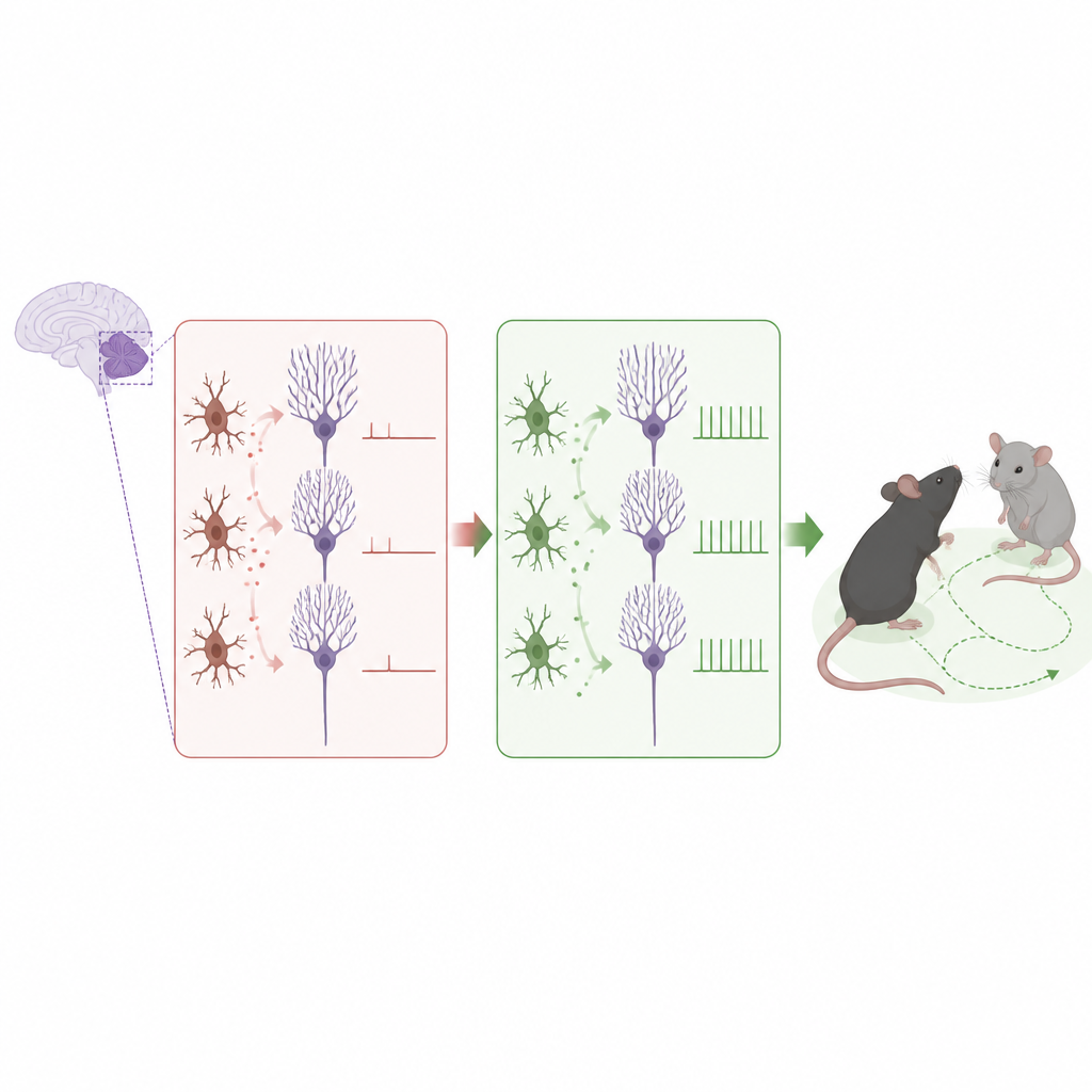
The team then asked what IL-17A actually does to these cells and to behavior. Adding IL-17A to cerebellar slices increased the spontaneous firing of Purkinje cells and reduced the strength of inhibitory signals they received, without changing excitatory input. When IL-17A was injected into Crus I of healthy mice, the animals spent more time engaging with an unfamiliar mouse, but their low baseline levels of repetitive behavior did not change. In contrast, blocking the IL-17 receptor or lowering its levels specifically in Purkinje cells made normally behaving mice show reduced sociability and more repetitive digging. These experiments indicate that a certain amount of IL-17A signaling is needed to keep Purkinje cells active enough to support healthy social interactions.
Rescuing autism-like traits through targeted immune signals
In Fragile X mice, IL-17A levels and IL-17RA receptors in the cerebellum were already higher than normal, suggesting a built-in attempt to compensate for circuit problems. Boosting this pathway further had striking effects. Direct IL-17A delivery to Crus I restored Purkinje cell firing to near-normal levels, weakened overly strong inhibitory contacts, and reduced the abundance of key inhibitory proteins at these synapses. Behaviorally, treated mice became more sociable and showed fewer repetitive actions. The researchers also tested a viral mimic called poly(I:C), used clinically in cancer immunotherapy, which gently stimulated microglia to release more IL-17A in the cerebellum. Poly(I:C) improved social preference and reduced repetitive behavior in both male and female Fragile X mice. These benefits disappeared if microglia were depleted or if IL-17A signaling in Crus I was blocked, confirming that local microglia-to-Purkinje communication was essential.
What this means for autism and the fever effect
Together, the findings reveal that IL-17A made by cerebellar microglia is not just an inflammatory trigger but a local tuner of brain circuits that govern social behavior. In the Fragile X model, raising IL-17A levels or enhancing its release acts as a compensatory mechanism that helps correct underactive Purkinje cells and eases autism-like symptoms, without obvious damage to neurons. This work offers a possible explanation for the so-called fever effect, where some individuals with autism temporarily improve during infections that activate the immune system. It also opens the door to therapies that carefully harness immune signals within specific brain regions to rebalance activity in social circuits, while avoiding broad immune activation.
Citation: Yin, J., Li, W., Shen, LP. et al. Cerebellar microglia-derived IL-17A mitigates autism-related behavioral and synaptic deficits. Mol Psychiatry 31, 3154–3168 (2026). https://doi.org/10.1038/s41380-026-03454-1
Keywords: autism, cerebellum, microglia, IL-17A, social behavior