Clear Sky Science · en
Multilayered microelectrode array for monitoring electrophysiological signals of 3d neural networks in cerebral organoid
Listening to Tiny Brains in Three Dimensions
Scientists are learning to grow miniature, brain-like tissues in the lab, known as cerebral organoids. These living models can mimic some features of the human brain and may help us understand disorders, test drugs, and explore new forms of computing. But to make the most of them, researchers need better ways to listen to the electrical chatter of their nerve cells deep inside, not just on the surface. This study introduces a new device that can record signals from multiple depths inside these tiny brains without cutting or damaging them.
A Gentle Scaffold for Growing Mini Brains
Organoids are soft spheres of living cells, while most electronics are flat and rigid. The team tackled this mismatch by building a flexible scaffold made of thin, porous films with tiny metal spots that act like microphones for nerve cells. These films are stacked with soft spacers between them, creating several layers that an organoid can grow into. The large pores let cells weave through the structure and allow nutrients and oxygen to flow freely, helping the tissue stay healthy over time. This design turns the device into both a support frame and a listening platform for activity throughout the three-dimensional tissue. 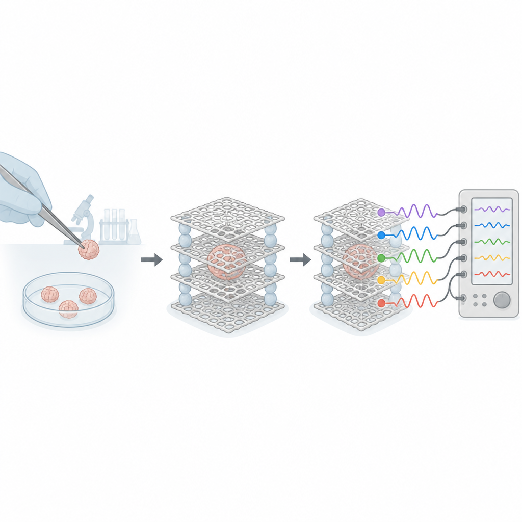
Custom Layers for Different Experiments
The researchers showed that the distance between layers can be finely tuned by changing the thickness of the soft spacers. They used imaging to confirm that the layers stay well aligned and that the gaps between them match the intended values. The mesh films are strong enough to be handled repeatedly yet thin enough to bend, which means they can be arranged in flat stacks, gentle curves, or more complex shapes. The team even demonstrated versions with four layers and layouts that can host several organoids at once, opening the door to high-throughput studies or side-by-side tests of different treatments on multiple samples.
Stable Signals from Deep Inside the Tissue
To pick up faint electrical spikes from neurons, the team coated each tiny electrode with a rough layer of platinum that lowers electrical resistance and improves signal quality. They used computer simulations to check that the structure would not sag or deform under the small weight of an organoid, and found that the spacers help keep strains low and spacing stable. They then grew cerebral organoids from human stem cells, let them mature, and gently placed them on the top mesh. Over several weeks, the organoids thickened and gradually infiltrated deeper layers, all while maintaining healthy cell markers and strong contact with the porous scaffold.
Following Neural Conversations in 3D
Using their multilayer device, the researchers recorded electrical activity from two and then four layers at once as the organoids developed. Early on, the neurons fired occasional, scattered spikes. With time, the signals became more frequent and more synchronized, forming bursts that appeared across multiple depths. The fraction of active recording sites grew steadily, and the quality of the signals remained high, showing that the device stayed well coupled to the tissue. By analyzing the timing of spikes across electrodes, the team built three-dimensional maps of how different regions of the organoid communicated, revealing evolving patterns of connectivity and coordinated activity between layers. 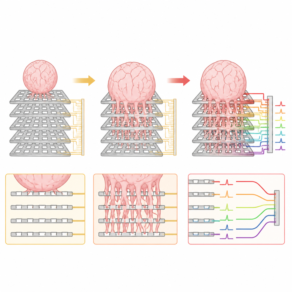
Poking the Network and Probing Its Limits
The device is not only a passive listener. In later experiments, the researchers delivered small, carefully chosen electrical pulses through one part of the array and watched how the organoid responded. The stimulation triggered both local and widespread changes in activity and increased the coordination between different sites, indicating that the network could be driven and reshaped by external input. The authors also discuss current limitations, such as difficulty pinpointing the exact position of each electrode inside the organoid and the natural variability in how organoids grow and spread across the mesh. They outline future improvements, including better shaping of the scaffold and combining electrical recordings with advanced imaging.
What This Means for Future Brain Research
In plain terms, this work shows a way to hear the electrical signals of a tiny, growing brain-like tissue in three dimensions without slicing it open. The multilayer mesh system lets scientists track how networks of nerve cells form, change, and respond to stimulation throughout the volume of an organoid. This approach could make organoids more useful for studying brain development, disease processes, and the effects of medicines, and may even support new kinds of bio-based computing. While there is still work to do to better map exact locations and long-term effects, the device offers a promising bridge between flat electronics and the complex, layered structure of living brain tissue.
Citation: Kim, N., Kang, M., Ji, J. et al. Multilayered microelectrode array for monitoring electrophysiological signals of 3d neural networks in cerebral organoid. Microsyst Nanoeng 12, 201 (2026). https://doi.org/10.1038/s41378-026-01328-8
Keywords: cerebral organoids, microelectrode arrays, 3D neural networks, electrophysiology, brain models