Clear Sky Science · en
UNIQUE: ultrasound non-destructive in-situ quantitative evaluation of stem cell spheroid deformability during differentiation into specific lineages
Why gentle tests on tiny cell clusters matter
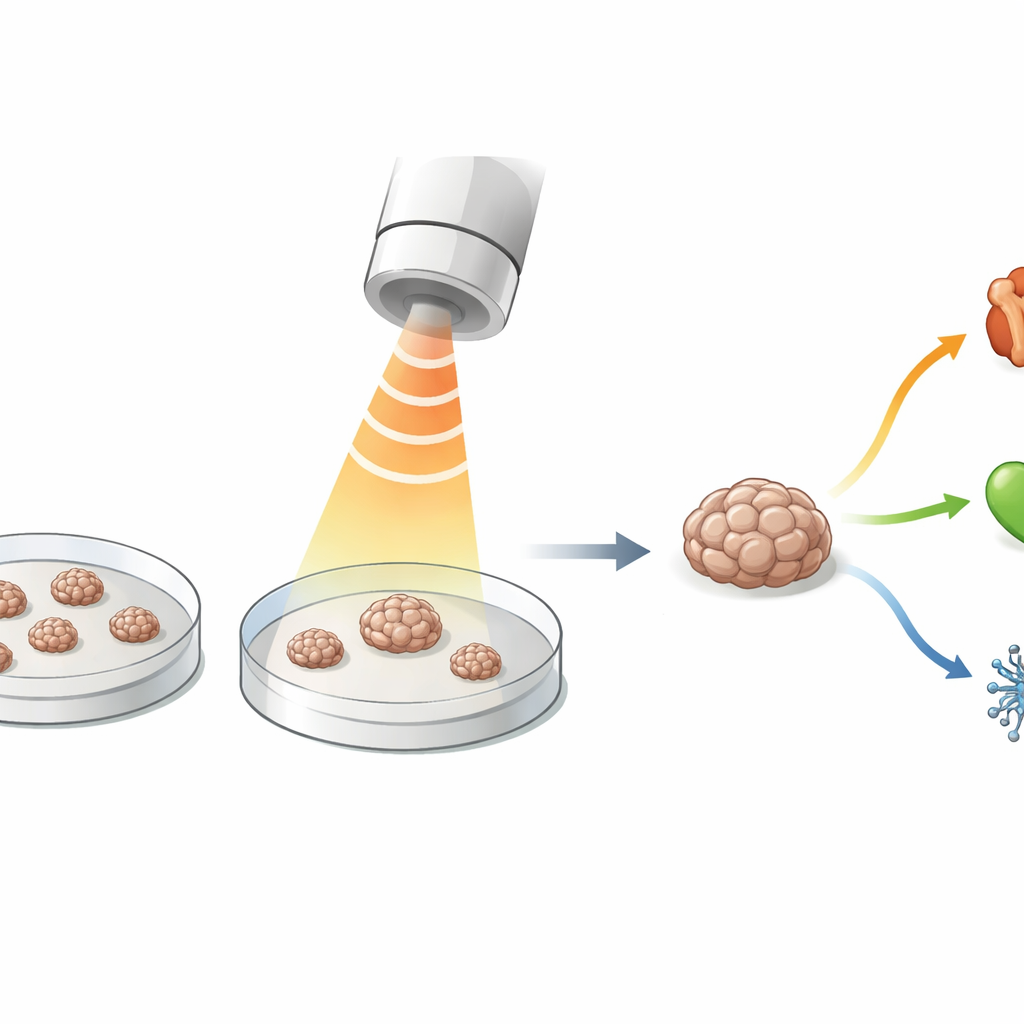
Doctors hope to repair worn joints, damaged hearts, and other tissues using living cells grown in the lab. A promising approach uses tiny three-dimensional clusters of stem cells, called spheroids, which survive and function better than loose cells when transplanted. But today, checking whether each spheroid is healthy and maturing into the right tissue usually destroys it. This study introduces a gentle ultrasound-based method that can probe the softness of individual spheroids in their normal culture dish and follow their development over time without harming them.
Soft sound waves as a new testing tool
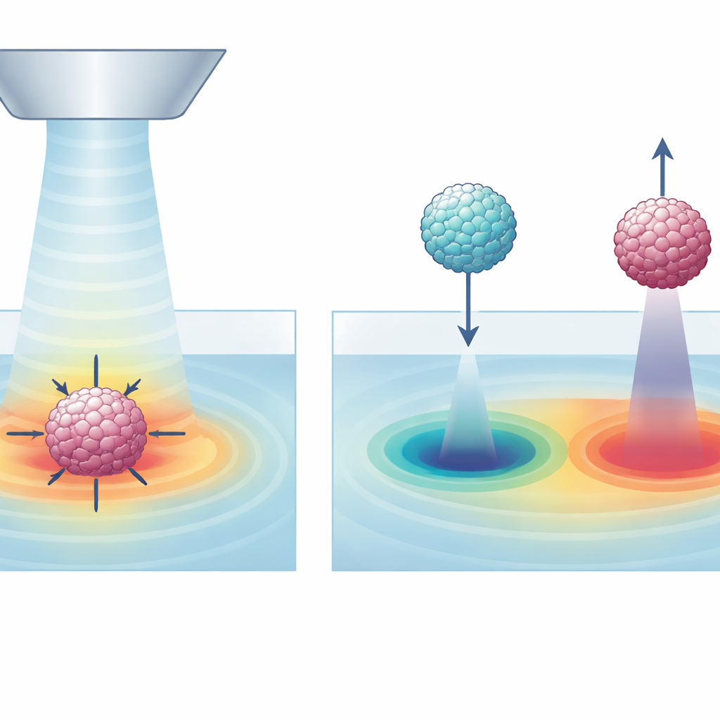
The researchers created a platform they call UNIQUE that uses focused ultrasound to push very lightly on a single stem cell spheroid. A small ultrasound beam is aimed at the spheroid inside an ordinary culture dish, causing the cluster to deform slightly. High-resolution microscopes record how much its cross-sectional area changes. From this tiny shape change, the team calculates a measure of deformability, which reflects how soft or stiff the spheroid is. By tuning the strength, timing, and position of the ultrasound, they found settings that produce clear, repeatable measurements while keeping the spheroids intact and viable.
Linking softness to future cell identity
To show what this mechanical test can reveal, the team worked with human adipose-derived stem cells, a versatile type of cell taken from fat tissue. They formed these cells into spheroids and then guided them toward three common fates: fat-like, cartilage-like, and bone-like tissues. Using UNIQUE, they measured deformability of individual spheroids every few days during the three-week maturation period and compared these readings with standard molecular markers. Fat-forming spheroids became progressively softer, while cartilage-forming and especially bone-forming spheroids tended to stiffen or change less, matching known changes in their internal structure and surrounding matrix.
Using feel to predict where cells are headed
Because stem cells from different donors behave differently, the authors also asked whether early mechanical readings could forecast how well spheroids would later mature. They found that initial deformability varied among donors in a stable way and that these early values correlated with how strongly the spheroids later expressed lineage markers. In simple terms, softer starting spheroids were more likely to show robust fat development, and greater deformability generally aligned with more advanced stages of all three tissue types. This suggests that a quick mechanical check before differentiation begins could help select spheroids with better therapeutic potential.
Sorting spheroids by how they respond to sound
Beyond measuring deformability, UNIQUE can also act as a kind of acoustic tweezer. The focused ultrasound field creates regions of low and high acoustic energy that either pull spheroids toward the beam center or push them away, depending on their internal makeup. The team showed that fat-forming spheroids, which develop many lipid droplets and change their acoustic properties, are drawn into the focal region and can be stably held and moved in two dimensions. In contrast, cartilage- and bone-forming spheroids tend to be repelled. This behavior allows label-free sorting of spheroids based purely on their mechanical and acoustic character, while measurements continue in real time.
What this means for future cell therapies
The UNIQUE platform shows that gentle ultrasound can monitor how tiny stem cell clusters soften or stiffen as they turn into specific tissues and that these mechanical fingerprints can hint at their future behavior. Because the method works in standard culture dishes, does not require fluorescent tags, and does not damage the samples, it could become a practical quality-control tool for manufacturing cell-based therapies. By giving researchers and clinicians a way to continuously watch the physical state of living spheroids, this technology may help ensure that only well-formed, correctly committed cell clusters are chosen for regenerative treatments.
Citation: Ha, H., Yoo, J., Kang, Y. et al. UNIQUE: ultrasound non-destructive in-situ quantitative evaluation of stem cell spheroid deformability during differentiation into specific lineages. Microsyst Nanoeng 12, 166 (2026). https://doi.org/10.1038/s41378-026-01305-1
Keywords: stem cell spheroids, ultrasound, cell mechanics, regenerative medicine, non-destructive evaluation